Sensing the World Through Whiskers
- Published22 Aug 2014
- Reviewed22 Aug 2014
- Author Summer Allen
- Source BrainFacts/SfN
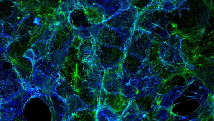
Through our senses, we experience the world. For rodents, the facial whiskers act as a channel through which they obtain information about their surroundings. By studying whiskers in rodents, scientists hope to gain general insight into how the brain processes sensory experiences.
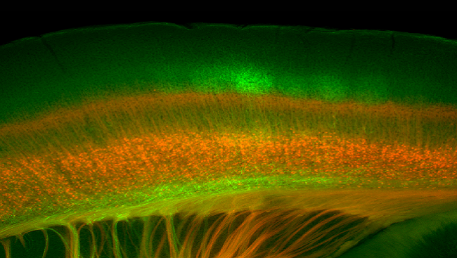
This image of a mouse brain shows the endings of nerve cells that transport sensory information (in green) from the thalamus, the brain’s sensory relay station, to the barrel cortex, a part of the brain that processes whisker information. The neurons in red send information back to the thalamus to control the way the brain responds to the whisker sensations. Learning how these cells work together may one day contribute to researchers' understanding of normal and abnormal sensory perception.
About the Author

Summer Allen is a science writer and postdoctoral researcher at Brown University. While her research focuses on genes that control how neurons communicate, she enjoys learning about all topics in neuroscience.
CONTENT PROVIDED BY
BrainFacts/SfN