Show Some Spine
- Published12 Mar 2019
- Reviewed12 Mar 2019
- Author Michael W. Richardson
- Source BrainFacts/SfN
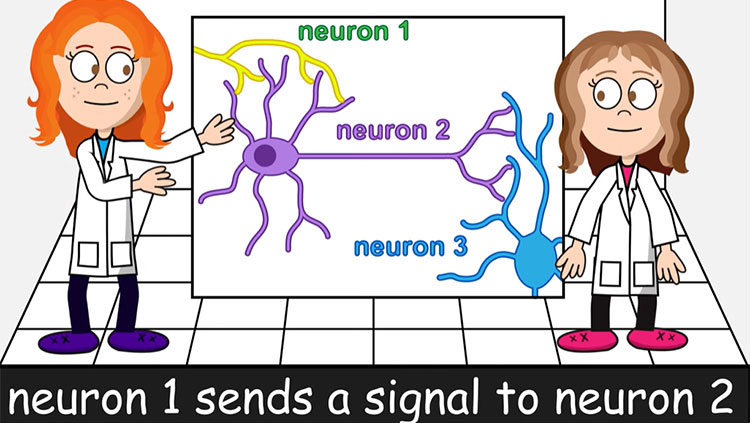
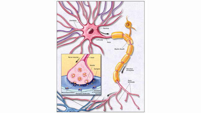
Your brain is just one part of your nervous system. The peripheral nervous system, encompassing parts like motor neurons (responsible for muscle movement) and the nerves behind your sense of touch, spreads like a river delta throughout your body. When signals need to travel back and forth to the brain, they move along the superhighway that is the spinal cord.
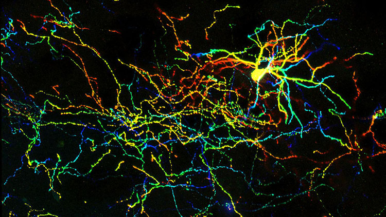
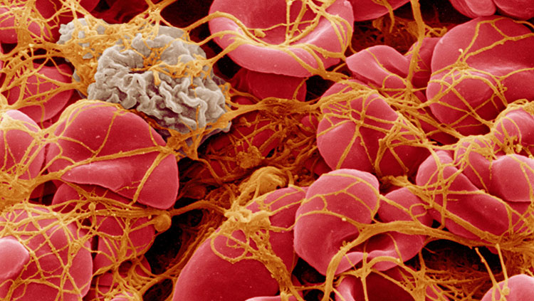
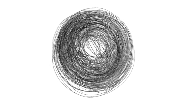
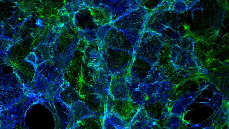
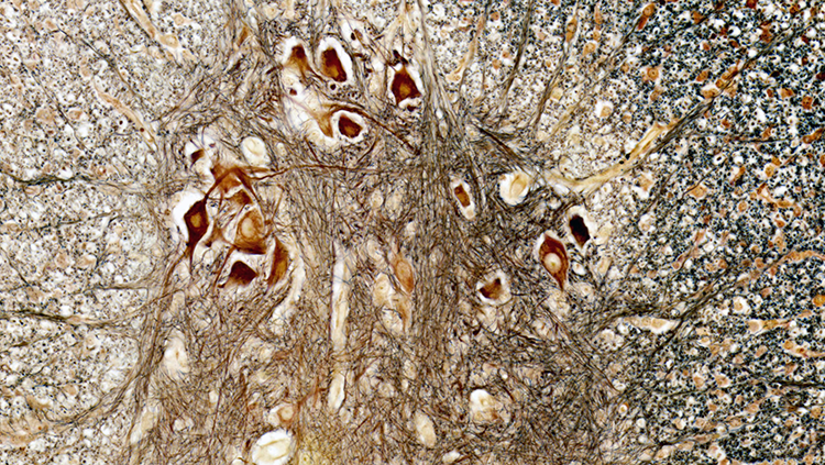
Made of neural tissue and partly enclosed by vertebrae, the spinal cord stretches from the medulla oblongata to your lower back and contains bundles of axons that connect everything from your brain down to your toes. In this cross view of the spinal cord, large nerve cells running up the spine can be seen in brown and yellow. They are surrounded by smaller dendrites and nerves, delivering signals up and down the spine.
About the Author

Michael W. Richardson is a writer and editor based in Brooklyn, New York, covering topics ranging from the brain and behavior to the environment.
CONTENT PROVIDED BY
BrainFacts/SfN