Magnetoencephalography
- Published1 Apr 2012
- Reviewed1 Apr 2012
- Source BrainFacts/SfN
Much faster than a traditional MRI, MEG can be used to provide even more information about brain activity.
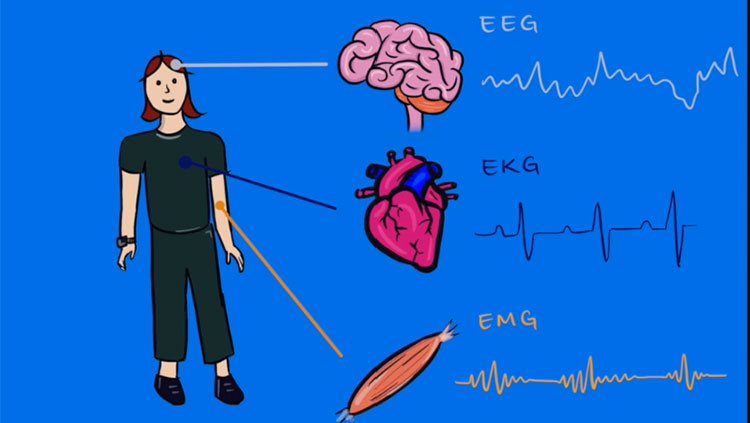
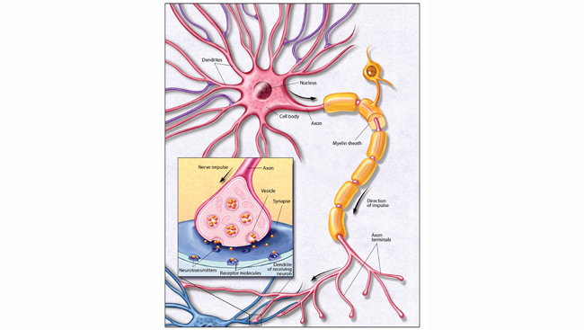
MEG is a recently developed technique that reveals the source of weak magnetic fields emitted by neurons. An array of cylinder-shaped sensors monitors the magnetic field pattern near the patient’s head to determine the position and strength of activity in various regions of the brain. In contrast with other imaging techniques, MEG can characterize rapidly changing patterns of neural activity — down to millisecond resolution — and can provide a quantitative measure of the strength of this activity in individual subjects. Moreover, by presenting stimuli at various rates, scientists can determine how long neural activation is sustained in the diverse brain areas that typically respond.
One of the most exciting developments in imaging is the combined use of information from fMRI and MEG. The former provides detailed information about the areas of brain activity while an individual is engaged in a particular task, whereas MEG tells researchers and physicians when certain areas become active. Together, this information leads to a much more precise understanding of how the brain works in health and disease.
CONTENT PROVIDED BY
BrainFacts/SfN