Visualizing the Developing Brain
- Published27 Dec 2013
- Reviewed27 Dec 2013
- Author Michael W. Richardson
- Source BrainFacts/SfN
Gordon, et al. Journal of Neuroscience, 2013.
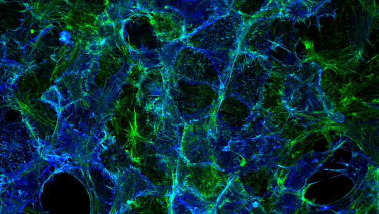
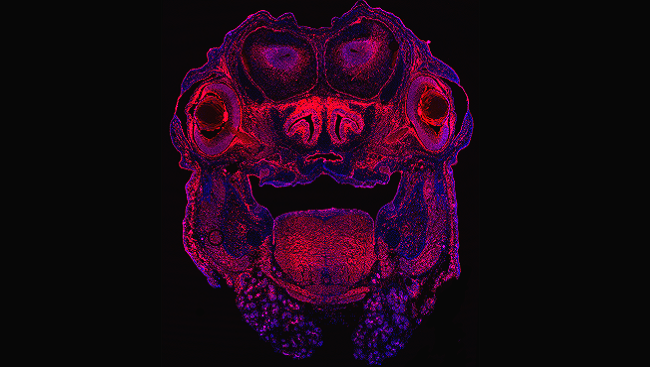
The brain is the body’s most complex organ — made up of more than 100 billion cells — so neuroscientists need a way to locate the particular cells they wish to study. Fluorescent labelling is one such technique. This image displays a cross-section of a mouse brain early in development, with the nuclei of nerve cells labeled in blue. In this study, scientists manipulated genes in stem and progenitor cells (appearing in red) to turn on or off so they could track their location and determine gene function in the developing brain.
About the Author

Michael W. Richardson is a writer and editor based in Brooklyn, New York, covering topics ranging from the brain and behavior to the environment.
CONTENT PROVIDED BY
BrainFacts/SfN