Keep in Touch
- Published26 Dec 2014
- Reviewed22 Dec 2014
- Author Alexis Wnuk
- Source BrainFacts/SfN
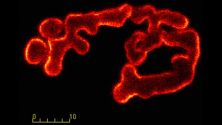
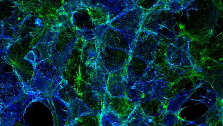
Whether you’re playing a complex concerto or simply walking, your brain must signal to your muscles to move. Nerve cells connect with muscle fibers to relay these messages at a point called the neuromuscular junction (pictured above). The image shows a neuromuscular junction in a mouse, where motor neurons (green) release the neurotransmitter acetylcholine, which signals the muscle fibers (red) to contract.
Neuromuscular diseases like congenital myasthenia and myasthenia gravis result from faulty connections between motor neurons and muscles. By studying the neuromuscular junction, scientists hope to gain insight into what goes wrong in these diseases.
About the Author

Alexis is a former staff writer/editor for BrainFacts.org. She graduated from the University of Pittsburgh in 2012 with degrees in neuroscience and English.
CONTENT PROVIDED BY
BrainFacts/SfN