A Visual Arrangement
- Published29 Sep 2015
- Reviewed28 Sep 2015
- Author Michael W. Richardson
- Source BrainFacts/SfN
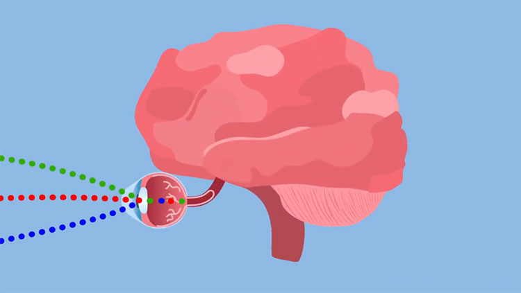
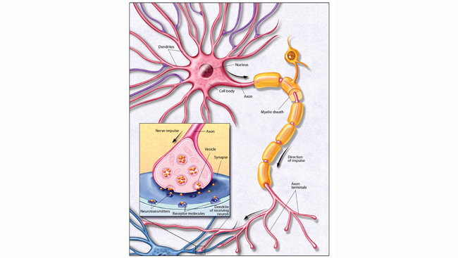
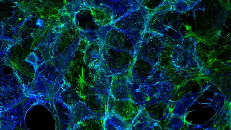
Cells in the retina are arranged on a flat plane, a bit like the spokes of a bicycle wheel. During development, their axons — the fiber-like extensions from neurons — grow toward the center of the retina. Here they join together to form the optic nerve, the superhighway that carries visual information from the eye to the brain.
However, problems during development can cause the axons to grow in abnormal directions rather than toward the center of the retina, and this can lead to visual impairment.
This image shows the retinas of two embryonic mice. The retina on the left is healthy; the axons (purple) are all arranged on the same plane and growing toward the center. The retina on the right, however, has axons (red and yellow) that are extending in abnormal directions, invading other parts of the eye. By studying what guides axons on the correct path, scientists hope to better understand how the visual system develops and functions.
About the Author

Michael W. Richardson is a writer and editor based in Brooklyn, New York, covering topics ranging from the brain and behavior to the environment.
CONTENT PROVIDED BY
BrainFacts/SfN