Mapping the Brain
- Published1 Apr 2012
- Reviewed1 Apr 2012
- Source BrainFacts/SfN
The cerebrum, the largest part of the human brain, is associated with higher order functioning, including the control of voluntary behavior. Thinking, perceiving, planning, and understanding language all lie within the cerebrum’s control.
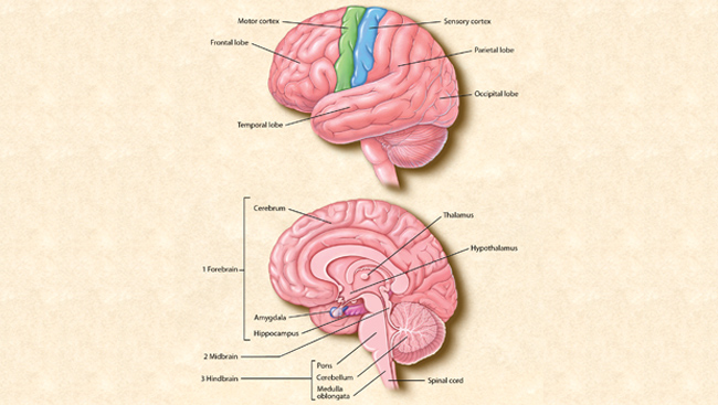
The top image shows the four main sections of the cerebral cortex: the frontal lobe, the parietal lobe, the occipital lobe, and the temporal lobe. Functions such as movement are controlled by the motor cortex, and the sensory cortex receives information on vision, hearing, speech, and other senses. The bottom image shows the location of the brain's major internal structures.
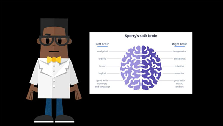
The cerebrum is divided into two hemispheres — the right hemisphere and the left hemisphere. Bridging the two hemispheres is a bundle of fibers called the corpus callosum. The two hemispheres communicate with one another across the corpus callosum.
Covering the outermost layer of the cerebrum is a sheet of tissue called the cerebral cortex. Because of its gray color, the cerebral cortex is often referred to as gray matter. The wrinkled appearance of the human brain also can be attributed to characteristics of the cerebral cortex. More than two-thirds of this layer is folded into grooves. The grooves increase the brain’s surface area, allowing for inclusion of many more neurons.
The function of the cerebral cortex can be understood by dividing it somewhat arbitrarily into zones, much like the geographical arrangement of continents.
The frontal lobe is responsible for initiating and coordinating motor movements; higher cognitive skills, such as problem solving, thinking, planning, and organizing; and for many aspects of personality and emotional makeup.
The parietal lobe is involved with sensory processes, attention, and language. Damage to the right side of the parietal lobe can result in difficulty navigating spaces, even familiar ones. If the left side is injured, the ability to understand spoken and/or written language may be impaired.
The occipital lobe helps process visual information, including recognition of shapes and colors.
The temporal lobe helps process auditory information and integrate information from the other senses. Neuroscientists also believe that the temporal lobe has a role to play in short-term memory through its hippocampal formation, and in learned emotional responses through its amygdala.
All of these structures make up the forebrain. Other key parts of the forebrain include the basal ganglia, which are cerebral nuclei deep in the cerebral cortex; the thalamus; and the hypothalamus. The cerebral nuclei help coordinate muscle movements and reward useful behaviors; the thalamus passes most sensory information on to the cerebral cortex after helping to prioritize it; and the hypothalamus is the control center for appetites, defensive and reproductive behaviors, and sleep-wakefulness.
The midbrain consists of two pairs of small hills called colliculi. These collections of neurons play a critical role in visual and auditory reflexes and in relaying this type of information to the thalamus. The midbrain also has clusters of neurons that regulate activity in widespread parts of the central nervous system and are thought to be important for reward mechanisms and mood.
The hindbrain includes the pons and the medulla oblongata, which control respiration, heart rhythms, and blood glucose levels.
Another part of the hindbrain is the cerebellum which, like the cerebrum, also has two hemispheres. The cerebellum’s two hemispheres help control movement and cognitive processes that require precise timing, and also play an important role in Pavlovian learning.
The spinal cord is the extension of the brain through the vertebral column. It receives sensory information from all parts of the body below the head. It uses this information for reflex responses to pain, for example, and it also relays the sensory information to the brain and its cerebral cortex. In addition, the spinal cord generates nerve impulses in nerves that control the muscles and the viscera, both through reflex activities and through voluntary commands from the cerebrum.
CONTENT PROVIDED BY
BrainFacts/SfN
Also In Anatomy

Trending
Popular articles on BrainFacts.org