Myelin: An Overview
- Published24 Mar 2015
- Reviewed24 Mar 2015
- Author Caitlin Kirkwood
- Source BrainFacts/SfN
Myelin is a fatty substance that wraps around nerve fibers and serves to increase the speed of electrical communication between neurons. While the function of myelin remained elusive for many years, today scientists are putting their knowledge about this insulating substance to use and investigating strategies to protect and repair myelin in diseases where it is compromised like multiple sclerosis.
The Nervous System’s Insulation
Communication between neurons depends on the spread of electrical signals, and, just as wires need to be insulated, so too do neurons. Myelin was discovered in the mid-1800s, but nearly half a century passed before scientists discovered its vital role as an insulator.
Myelin’s Discovery
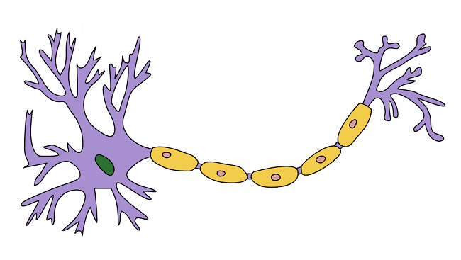
In the mid-19th century, scientists peering into light microscopes noticed something strange about the nerve fibers (axons) branching from the spinal cord: they were surrounded by a glistening, white, fatty substance. While making this observation, German pathologist Rudolf Virchow coined the term “myelin,” from the Greek word myelós, meaning marrow or core. At the time he thought that myelin was present inside of the nerve fiber and incorrectly likened the substance to bone marrow. Made of lipids and proteins, myelin was later found to wrap around the axons of neurons.
Myelin is made by two different types of support cells. In the central nervous system (CNS) — the brain and spinal cord — cells called oligodendrocytes wrap their branch-like extensions around axons to create a myelin sheath. In the nerves outside of the spinal cord, Schwann cells produce myelin. Regardless of where it is in the nervous system, all myelin performs the same function, enabling efficient transmission of electrical signals.
Speeding Up Neural Communication
Even the best insulation would have trouble maintaining electrical signals over long distances, and axons in large animals like giraffes can be up to 15 feet long. Research through the 1940s showed how myelin ensures that the signal is maintained and transmitted.
In the 1870s, French physician Louis-Antoine Ranvier noted that the myelin sheath is discontinuous, covering most of the nerve fiber but with gaps at regular intervals along the axon. Scientists later learned that charged particles called ions can cross the axon only at these myelin gaps, which became known as the “nodes of Ranvier.”
In the 1930s and 1940s, scientists found that this passage of ions helps maintain the electrical signal, allowing it to travel quickly down an axon. The signal appears to “jump” from one node to the next in a process called saltatory conduction.
Insight Into Myelin’s Role in Health and Disease
Removing Myelin Disrupts Neural Communication
With knowledge of myelin’s role in neural communication, researchers aimed to find out what happens when myelin is disrupted. In the 1980s, researchers used animal models to assess how electrical nerve signals are altered after axons were stripped of the myelin (demyelinated). When researchers chemically induced myelin loss in the spinal cords of cats, they found that signals moved more slowly along the nerve fiber and often failed to make it to the end of the axon.
Around the same time, scientists also made breakthroughs in identifying many of the components of myelin, like the major protein elements of the myelin sheath and the genes that encode them. Researchers developed mouse models that had defective myelin proteins, resulting in a myelin deficiency. One such mouse is the “shiverer” mouse, named after its body tremors. Mice like the shiverer mouse have provided researchers with a model system for studying myelin’s function in the healthy nervous system and its dysfunction in demyelinating diseases.
Myelin Loss in Disease
Loss of myelin is a problem for many CNS disorders, including stroke, spinal cord injury, and, most notably, multiple sclerosis (MS).
MS is a chronic, disabling disease of the CNS that affects more than 2.3 million people worldwide. MS results from the accumulation of damage to myelin and the underlying nerve fibers it insulates and protects.
Current research indicates that MS involves an autoimmune response. Scientists think that immune cells, which normally defend the body against bacteria and viruses, mistakenly attack the myelin sheath, stripping it away and exposing the nerve fibers underneath. In addition, recent research suggests that axon damage occurs early on in the course of the disease. Once damaged, the ability of nerve cells in the brain and spinal cord to communicate with each other and with muscles is compromised, leading to a variety of unpredictable symptoms that vary from person to person. These symptoms, which can be temporary or permanent, range from fatigue, weakness, and numbness to blindness and even paralysis.
New Treatment Possibilities for Demyelinating Diseases
Research understanding the components of myelin, how it is produced, and how it functions has paved the way for new therapeutic possibilities in myelin-degenerative diseases like MS.
Repair and Protect
Repairing and protecting myelin is one of the approaches to treating demyelinating disease like MS. This approach focuses on (1) repairing the damage that has already occurred and (2) preventing further injury to nerves and axons.
Several drugs that are currently approved for treating MS follow the second strategy. They work by suppressing or changing the activity of the immune system, protecting myelin from unwarranted attacks. However, to date none of the available medications address regeneration of lost myelin.
Scientists are exploring many lines of research to repair and protect myelin, including developing therapies that stimulate the brain’s natural capacity to heal itself, discovering new drug targets for myelin regeneration, and piloting stem cell therapies in animal models.
Stem Cells’ Promise
Stem cell therapy is one avenue being explored in the search for treatments for MS. Scientists in the United States and Italy have taken skin cells from mice and “reprogrammed” them into stem cells that can become any kind of cell in the nervous system. These new stem cells were then infused into the spinal cords of mice models of MS where they secreted factors that helped the myelin-producing cells survive. Consequently, these mice had more myelination and less axonal damage compared to mice that did not receive stem cell infusions. While the results are promising, much more work will need to be done in human clinical trials to determine the therapeutic efficacy.
Myelin’s discovery more than a century ago has advanced our understanding of how the nervous system functions. Continued research efforts funded by public and private institutions worldwide seek to understand how myelin is compromised in diseases like MS, revealing new possibilities for treatment and offering hope to the millions of people affected by these diseases.
About the Author

Caitlin Kirkwood is a freelance science writer and PhD candidate in neurobiology at the University of Pittsburgh. Her translational research focuses on molecular mechanisms of Alzheimer's disease and psychosis. In addition to contributing to BrainFacts.org, she has written for Scientific American and Being Human.
CONTENT PROVIDED BY
BrainFacts/SfN
References
Deoni SCL, Mercure E, Blasi A, Gasston D, Thomson A, et al. Mapping infant brain myelination with magnetic resonance imaging. Journal of Neuroscience. 31(2): 784-791 (2011).
Gasser HS, Grundfest H. Axon diameters in relation to the spike dimensions and the conduction velocity in mammalian fibers. American Journal of Physiology. 127: 393-414 (1939).
Giedd JN. Structural magnetic resonance imaging of the adolescent brain. Annals of the New York Academy of Sciences. 1021: 77-85 (2004).
Huxley AF, Stampfli R. Evidence for saltatory conduction in peripheral myelinated nerve fibres. Journal of Physiology. 108: 315-339 (1949)
Laterza C, Merlini A, De Feo D, Ruffini F, Menon R, et al. iPSC-derived neural precursors exert a neuroprotective role in immune-mediated demyelination via the secretion of LIF. Nature Communications. 4: 1-16 (2013).
Miller DJ, Duka T, Stimpson CD, Schapiro SJ, Baze WB, et al. Prolonged myelination in human neocortical evolution. Proceedings of the National Academy of Sciences. 109(41): 16480-16485 (2012).
Roach A, Boylan K, Horvath S, Rusiner SB, Hood LE. Characterization of cloned cDNA representing rat myelin basic protein: absence of expression in brain of shiverer mutant mice. Cell. 34(3): 799-806 (1983).
Rushton WAH. A theory of the effects of fibre size in medullated nerve. Journal of Physiology. 115(1): 101-122 (1951).
Sampaio-Baptista C, Khrapitchev AA, Foxley S, Schlagheck T, Scholz J, et al. Motor Skill Learning Induces Changes in White Matter Microstructure and Myelination. Journal of Neuroscience. 33(50): 19499-19503 (2013).
Smith KJ, Blakemore WF, McDonald WI. The restoration of conduction by central remyelination. Brain. 104: 383-404 (1981).
Tasaki I. The electro-saltatory transmission of the nerve impulse and the effect of narcosis upon the nerve fiber. American Journal of Physiology. 127: 211-227 (1939).
Waxman SG. Determinants of conduction velocity in myelinated nerve fibers. Muscle & Nerve. 3: 141-150 (1980).
Yakovlev PI, Lecours A. The myelogenetic cycles of regional maturation of the brain. In: Minkowski A, Regional development of the brain in early life. Blackwell, Oxford, 3–70 (1967).