Identifying Major Brain Landmarks
- Reviewed8 Nov 2022
- Author Diane A. Kelly
- Source BrainFacts/SfN
The brain is literally the “nerve center” of your body — it contains billions of neurons that transmit information from the body and the outside world, and then programs our responses — conscious and unconscious movements, thoughts, emotions, and memories. What’s more, your brain can do all these things simultaneously: You can throw a ball while talking to a friend, plan dinner while you’re shopping, or daydream about a balloon ride as you drive to work. Your brain can pull off these feats of multitasking because it is split into many distinct regions specialized for specific tasks and abilities.
The largest part of the human brain is the cerebrum. It is divided into two large, separate hemispheres: one on the left side and the other on the right. The hemispheres are connected by bundles of nerve fibers that carry information from one side of your brain to the other. The largest of these bundles forms a bridge between the cerebral hemispheres and is called the corpus callosum.
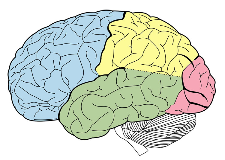
The surface of the cerebrum is a deeply folded layer of nerve tissue called the cerebral cortex. Its deep folds increase the area of the cerebral cortex, creating space in this surface layer for more neurons, which increase the brain’s processing power. Just as explorers use landmarks like rivers and mountain ranges to describe and map continents, neuroscientists use the deepest divisions of the cerebrum to identify regions of each hemisphere as separate lobes — distinct regions that have characteristic functions.
The frontal lobes are at the front of the brain, immediately above the eyes. Parts of these lobes coordinate voluntary movements and speech, memory and emotion, higher cognitive skills like planning and problem-solving, and many aspects of personality.
The parietal lobes are located at the top of the brain, immediately behind the frontal lobes. They integrate sensory signals from the skin, process taste, and process some types of visual information.
The back of the brain houses the occipital lobes. They process visual information and are responsible for recognizing colors and shapes, and integrating them into complex visual understanding.
The temporal lobes lie on the sides of the brain, at and below the level of the eyes. They carry out some visual processing and interpret auditory information. The hippocampus consists of curved structures lying beneath the cerebral cortex; it is a region of the temporal lobes that encodes new memories. Another deep structure within each temporal lobe, the amygdala, integrates memory and emotion.
The hippocampus and amygdala are part of the limbic system, a group of structures deep within the brain that help regulate our emotion and motivation. Other parts of the limbic system include the thalamus, which integrates sensory information and relays it to other parts of the brain, and the hypothalamus, which sends hormonal signals to the rest of the body through the pituitary gland. These structures, together with the cerebral cortex, make up the forebrain.
The midbrain sits beneath the thalamus. It includes distinct groups of neurons that coordinate eye movements like blinking and focusing, and trigger reflexes to sounds. An example is the startled jump when you are surprised by a loud noise. Other regions of the midbrain inhibit unwanted body movements and help coordinate sensory input and motor output to manage the fine motor control that enables you to write with a pen or play a musical instrument.
Some of these regions — along with parts of the forebrain — form a collection of structures called the basal ganglia, which helps regulate complex body movements.
The hindbrain plays roles in glucose regulation and sleep and includes several regions that help control movement. The cerebellum, tucked underneath the occipital lobe at the very back of the brain, is the second-largest part of the brain in volume, containing over half the brain’s neurons. Like the cerebrum, the cerebellum is deeply folded, divided into two hemispheres, and carries out a variety of functions. For example, it coordinates voluntary movements and helps the brain learn new motor skills. It also has roles in spatial and temporal perception. A patient with cerebellar damage might have a jerky, arrhythmic gait or might be unable to accurately touch his finger to his nose.
Below the cerebellum is the pons, which influences breathing and posture. Another part of the hindbrain, the medulla, carries nerve pathways connecting the brain to the spinal cord and contains neural networks that help control basic functions like swallowing, heart rate, and breathing. Together, the midbrain, pons, and medulla make up the brainstem.
Adapted from the 8th edition of Brain Facts by Diane A. Kelly.
About the Author

Diane A. Kelly studies neuroscience and anatomy at UMass Amherst, with a focus on mapping the circuits of the social behavior network. Her science writing has appeared in a variety of publications, including Gizmodo, Wired.com, FiveThirtyEight, and Muse. You can find her on Twitter @DianeAKelly.
CONTENT PROVIDED BY
BrainFacts/SfN
References
Albuixech-Crespo, B., López-Blanch, L., Burguera, D., Maeso, I., Sánchez-Arrones, L., et al. (2017). Molecular regionalization of the developing amphioxus neural tube challenges major partitions of the vertebrate brain. PLOS Biology, 15(4): e2001573. https://doi.org/10.1371/journal.pbio.2001573
Barton, R. A., & Venditti, C. (2014). Rapid Evolution of the Cerebellum in Humans and Other Great Apes. Current Biology, 24(20), 2440–2444. https://doi.org/10.1016/j.cub.2014.08.056
Bekkers, J. M. (2011). Pyramidal neurons. Current Biology, 21(24), PR975. https://doi.org/10.1016/j.cub.2011.10.037
Belkhiria, C., Driss, T., Habas, C., Jaafar, H., Guillevin, R., & de Marco, G. (2017). Exploration and Identification of Cortico-Cerebellar-Brainstem Closed Loop During a Motivational-Motor Task: an fMRI Study. The Cerebellum, 16, 326–339. https://doi.org/10.1007/s12311-016-0801-1
Bromfield, E. B., Cavazos, J. E., Sirven, J. I. (2006). An Introduction to Epilepsy, https://www.ncbi.nlm.nih.gov/books/NBK2508/
Carpenter, R., & Reddi, B. (2012). Neurophysiology: A Conceptual Approach, 5th edition. Hodder Arnold: London.
Castro, A., Becerra, M., Manso, M. J., & Anadón, R. (2015). Neuronal organization of the brain in the adult amphioxus (Branchiostoma lanceolatum): A study with acetylated tubulin immunohistochemistry. The Journal of Comparative Neurology, 523(15), 2211–2232. https://doi.org/10.1002/cne.23785
Clarke, L. E., & Barres, B. A. (2013). Emerging roles of astrocytes in neural circuit development. Nature Reviews Neuroscience, 14, 311–321. https://doi.org/10.1038/nrn3484
Fain, G. L., & O’Dell T. J. (2014). Molecular and Cellular Physiology of Neurons, 2nd edition. Harvard University Press: Cambridge.
Forger, N. G. (2016). Epigenetic mechanisms in sexual differentiation of the brain and behaviour. Philosophical Transactions of the Royal Society B: Biological Sciences, 371(1688), 20150114. https://doi.org/10.1098/rstb.2015.0114
Frohlich, F. (2016). Network Neuroscience, 1st edition. Academic Press: London.
Guo, J. U., Ma, D. K., Mo, H., Ball, M. P., Jang, M. H., Bonaguidi, M. A., Balazer, J. A., Eaves, H. L., Xie, B., Ford, E., Zhang, K., Ming, G. L., Gao, Y., & Song, H. (2011). Neuronal activity modifies the DNA methylation landscape in the adult brain. Nature Neuroscience, 14, 1345–1351. https://doi.org/10.1038/nn.2900
Hammond, C. (2014). Cellular and Molecular Neurophysiology, 4th edition. Academic Press.
Human Brain. (2017). Allen Brain Atlas. Allen Institute for Brain Science. https://human.brain-map.org/
Lee, A., Fakler, B., Kaczmarek, L. K., & Isom, L. L. (2014). More Than a Pore: Ion Channel Signaling Complexes. The Journal of Neuroscience, 34(46), 15159–15169. https://doi.org/10.1523/JNEUROSCI.3275-14.2014
Noback, C. R. et al (eds.). (2005). The Human Nervous System: Structure and Function, 6th edition. Humana Press: Totowa NJ.
O'Muircheartaigh, J., Keller, S. S., Barker, G. J., & Richardson, M. P. (2015). White Matter Connectivity of the Thalamus Delineates the Functional Architecture of Competing Thalamocortical Systems. Cerebral Cortex, 25(11), 4477–4489. https://doi.org/10.1093/cercor/bhv063
Peer, M., Nitzan, M., Bick, A. S., Levin, N., & Arzy, S. (2017). Evidence for Functional Networks within the Human Brain's White Matter. The Journal of Neuroscience, 37(27), 6394–6407. https://doi.org/10.1523/JNEUROSCI.3872-16.2017
Pyka, M., & Cheng, S. (2014). Pattern Association and Consolidation Emerges from Connectivity Properties between Cortex and Hippocampus. PLOS ONE, 9(1), e85016. https://doi.org/10.1371/journal.pone.0085016
Saladin, K. (2015). Anatomy & Physiology: The Unity of Form and Function, 7th edition. McGraw Hill: New York.
Schneider, G. E. (2014). Brain Structure and its Origins: in Development and in Evolution of Behavior and the Mind. MIT Press: Cambridge.
Sheng, M., Kim, E. (2011). The postsynaptic organization of synapses. Cold Spring Harbor Perspectives in Biology, 3(12), a005678. https://pubmed.ncbi.nlm.nih.gov/22046028
Sporns, O. (2013). Structure and function of complex brain networks. Dialogues in Clinical Neuroscience, 15(3), 247–262. https://doi.org/10.31887/DCNS.2013.15.3/osporns
Verberne, A. J., Sabetghadam, A., & Korim, W. S. (2014). Neural pathways that control the glucose counterregulatory response. Frontiers in Neuroscience, 8(38). https://doi.org/10.3389/fnins.2014.00038
Wells, R. B. (2005). Cortical Neurons and Circuits: A Tutorial Introduction. https://webpages.uidaho.edu/rwells/techdocs/Cortical%20Neurons%20and%20Circuits.pdf