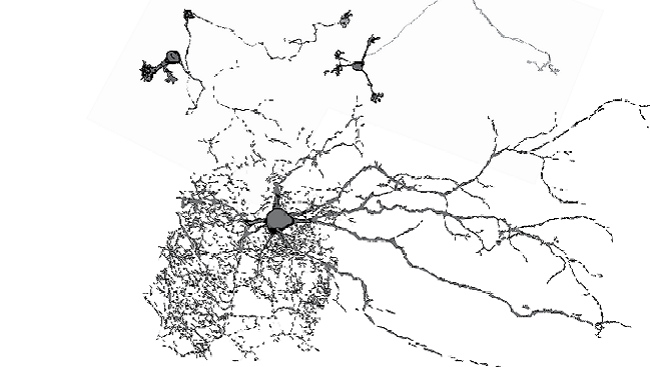
Researchers often study cells by isolating and filling them with chemicals to better visualize their structures. The image above shows different types of neurons in a mouse brain’s cerebellum that have been filled, scanned using imaging software, and reconstructed for more precise measurements.
Neurons are often named after their appearance. The tiny cell on the top right is called a cerebellar granule cell and the top left is a unipolar brush cell. The large one is called a Golgi cell, named for Camillo Golgi who won the Nobel Prize in 1906 .
The scientists who created this image studied receptors for the neurotransmitter gamma-aminobutyric acid (GABA), a chemical important in the regulation of sleep and sleep disorders.
CONTENT PROVIDED BY
BrainFacts/SfN