Forming the Sciatic Nerve
- Published7 Aug 2018
- Reviewed7 Aug 2018
- Author Charlie Wood
- Source BrainFacts/SfN
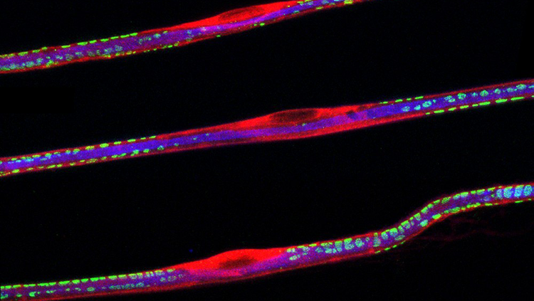
You might think of neurons as small (and they are, since these nerve fibers measure a fraction of a hair’s width across), but they come together to form mighty packages. These strands bundle up to form the largest nerve in your body — the sciatic nerve. It stretches from your lower back to the tip of your big toe, and matches the width of your thumb at its widest point. This sensation superhighway carries both feeling and movement signals up and down your legs, so doctors sometimes inject it with anesthesia during foot or leg surgery.
Here, the fibers’ neon glow comes from the different proteins that hold them together. The blue core highlights the filaments that form the neuron’s axons. The casings shine red from the Schwann cells that enclose and protect the axon, and the green spots represent a third protein embedded in the fiber.
CONTENT PROVIDED BY
BrainFacts/SfN
References
Charcot-Marie-Tooth Disease Fact Sheet. (n.d.). National Institute of Neurological Disorders and Stroke. Retrieved August 7, 2018, from https://www.ninds.nih.gov/Disorders/Patient-Caregiver-Education/Fact-Sheets/Charcot-Marie-Tooth-Disease-Fact-Sheet.
Lower Extremity Nerve Blocks. (n.d.). The New York School of General Anesthesia. Retrieved August 7, 2018, from http://www.nysora.com/files/2013/extremity-nerve-blocks/NYSORA_LowerExtremityProof10aFINAL.pdf
Praleema, R. R., MD. (n.d.). Anatomical Study of Width and Thickness of Sciatic Nerve in the Gluteal Region. World Research Library. Retrieved August 7, 2018, from http://www.worldresearchlibrary.org/up_proc/pdf/31-143229076226-28.pdf
Also In Cells & Circuits
Trending
Popular articles on BrainFacts.org


