Communicating Across Neural Networks
- Reviewed8 Nov 2022
- Author Diane A. Kelly
- Source BrainFacts/SfN
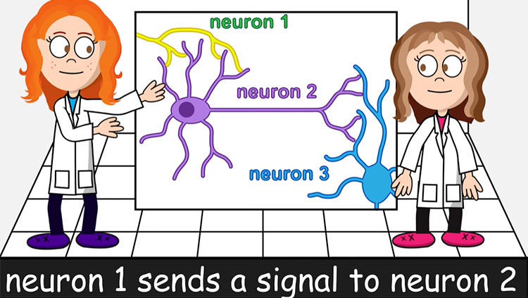
Although they have specialized functions, a brain region can communicate and work with other regions. Information moves from one region of your brain to another via chains of neurons that can transmit signals over long distances. When the nerve fibers of region-spanning neurons form distinct bundles, these are called nerve tracts. Examples of major nerve tracts include the corpus callosum (the thick bundle of neurons connecting your left and right cerebral hemispheres) and the smaller anterior commissure that transmits signals between the left and right temporal lobes.
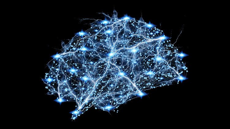
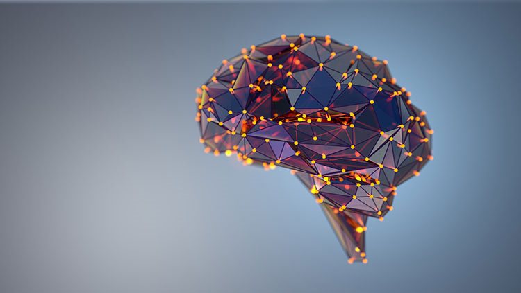
A group of nerve tracts connecting a series of regions in the brain is called a neural network. Neural net-works route signals through the brain along a linear pathway, analyzing and organizing different types of information within fractions of a second.
Have you ever wondered what happens in your brain when you watch a movie? Your brain turns a panoply of moving shapes into recognizable characters and scenery. The process begins with photoreceptors, cells in the retina that trigger electrical signals in response to specific wavelengths of light. Once those signals reach the optic nerve, they travel through the optic tract to the thalamus, where neurons respond to the shape, color, or movement of objects on the screen and pass their signals to the primary visual cortex in the occipital lobe at the back of the brain. Neurons in the primary visual cortex, in turn, detect the edges of objects within the field of vision and integrate the signals from each eye, creating a 3D representation of the outside world. The image is even further refined as signals are sent down two parallel processing streams. In one stream, neurons in the temporal lobe recognize and identify objects; in the other, neurons in the parietal lobe detect the spatial location of those objects. And that’s only the visual input from the film! New technologies that allow us to look with increasing detail at the brain regions being activated as we perform different functions are giving us increasing insight into the fine regions of the brain used for specific tasks.
Network Activity Creates Brain Waves
The visual cortex also sends signals back to the thalamus to become integrated with other sensory information; this is an example of a “thalamocortical loop,” a two-way circuit that connects the thalamus with parts of the cortex and back. As neuronal signals loop through the thalamus and cortex, they produce rhythmic, oscillating, electrical patterns that can be detected with an electroencephalograph (EEG). These signals are commonly called brain waves. There are four distinct types, each recognized by its characteristic shape on an EEG display or printout.
Your awake brain typically produces alpha waves and beta waves. Alpha waves originate mainly in the parietal and occipital lobes when your brain is relaxed and eyes are closed, and are characterized by frequencies between 8 and 13 Hertz (Hz). (The Hertz is a measure of frequency; 1 Hz = 1 cycle per second.) Beta waves are somewhat faster, with frequencies ranging from 14 to 30 Hz. Beta waves are typically produced by the frontal and parietal regions of your brain when it processes sensory input or concentrates on a task. Theta waves and delta waves are typical of sleep. Theta waves are slower than alpha waves, ranging from 4 to 7 Hz, while delta waves, which occur during deep sleep, are very slow, with frequencies less than 3.5 Hz. Alpha and delta waves are typically of higher amplitude (stronger) than beta or theta waves but, when measured with electrodes on your scalp, all these signals are in the microvolt range: 20–200 microvolts (μV) for alpha and delta waves, and 5-10 μV for beta and theta waves.
Neural Networks Organize and Integrate Information
Your brain and spinal cord contain many distinct neural networks. These include spinal tracts — chains of neurons that pass signals through the brainstem and the spinal cord. Signals either travel upward from sensory receptors in skin and muscles to the thalamus and parts of the cortex that interpret touch and pressure; or they travel downward from brain regions that induce movement, passing through the medulla and spinal cord before projecting to the body’s muscles.
Other neural networks provide feedback that helps integrate sensory and motor signals. For example, the brain’s basal ganglia are part of a feedback loop that takes information from cortical areas that elicit movement and produces signals that feed back to the cortex to excite or inhibit specific movements. Loops that connect the brainstem and the cerebellum also influence the timing and strength of motor signals; some of these loops incorporate tracts from the cerebral cortex that enable environmental and emotional context to influence your body’s movements. Networks that loop the hippocampus into sensory cortex pathways help your brain analyze whether environmental signals are familiar or are part of a new situation. Related networks linking the hippocampus to the thalamus and hypothalamus allow your memory to influence conscious behavior as well as unconscious physiological responses. Reflex loops are circuits eliciting action well before thoughts; these actions are controlled locally by information going in and out of the spinal cord or subcortical regions of the brain, and never reach the cortex.
Adapted from the 8th edition of Brain Facts by Diane A. Kelly.
About the Author

Diane A. Kelly studies neuroscience and anatomy at UMass Amherst, with a focus on mapping the circuits of the social behavior network. Her science writing has appeared in a variety of publications, including Gizmodo, Wired.com, FiveThirtyEight, and Muse. You can find her on Twitter @DianeAKelly.
CONTENT PROVIDED BY
BrainFacts/SfN
References
Albuixech-Crespo, B., López-Blanch, L., Burguera, D., Maeso, I., Sánchez-Arrones, L., et al. (2017). Molecular regionalization of the developing amphioxus neural tube challenges major partitions of the vertebrate brain. PLOS Biology, 15(4): e2001573. https://doi.org/10.1371/journal.pbio.2001573
Barton, R. A., & Venditti, C. (2014). Rapid Evolution of the Cerebellum in Humans and Other Great Apes. Current Biology, 24(20), 2440–2444. https://doi.org/10.1016/j.cub.2014.08.056
Bekkers, J. M. (2011). Pyramidal neurons. Current Biology, 21(24), PR975. https://doi.org/10.1016/j.cub.2011.10.037
Belkhiria, C., Driss, T., Habas, C., Jaafar, H., Guillevin, R., & de Marco, G. (2017). Exploration and Identification of Cortico-Cerebellar-Brainstem Closed Loop During a Motivational-Motor Task: an fMRI Study. The Cerebellum, 16, 326–339. https://doi.org/10.1007/s12311-016-0801-1
Bromfield, E. B., Cavazos, J. E., Sirven, J. I. (2006). An Introduction to Epilepsy, https://www.ncbi.nlm.nih.gov/books/NBK2508/
Carpenter, R., & Reddi, B. (2012). Neurophysiology: A Conceptual Approach, 5th edition. Hodder Arnold: London.
Castro, A., Becerra, M., Manso, M. J., & Anadón, R. (2015). Neuronal organization of the brain in the adult amphioxus (Branchiostoma lanceolatum): A study with acetylated tubulin immunohistochemistry. The Journal of Comparative Neurology, 523(15), 2211–2232. https://doi.org/10.1002/cne.23785
Clarke, L. E., & Barres, B. A. (2013). Emerging roles of astrocytes in neural circuit development. Nature Reviews Neuroscience, 14, 311–321. https://doi.org/10.1038/nrn3484
Fain, G. L., & O’Dell T. J. (2014). Molecular and Cellular Physiology of Neurons, 2nd edition. Harvard University Press: Cambridge.
Forger, N. G. (2016). Epigenetic mechanisms in sexual differentiation of the brain and behaviour. Philosophical Transactions of the Royal Society B: Biological Sciences, 371(1688), 20150114. https://doi.org/10.1098/rstb.2015.0114
Frohlich, F. (2016). Network Neuroscience, 1st edition. Academic Press: London.
Guo, J. U., Ma, D. K., Mo, H., Ball, M. P., Jang, M. H., Bonaguidi, M. A., Balazer, J. A., Eaves, H. L., Xie, B., Ford, E., Zhang, K., Ming, G. L., Gao, Y., & Song, H. (2011). Neuronal activity modifies the DNA methylation landscape in the adult brain. Nature Neuroscience, 14, 1345–1351. https://doi.org/10.1038/nn.2900
Hammond, C. (2014). Cellular and Molecular Neurophysiology, 4th edition. Academic Press.
Human Brain. (2017). Allen Brain Atlas. Allen Institute for Brain Science. https://human.brain-map.org/
Lee, A., Fakler, B., Kaczmarek, L. K., & Isom, L. L. (2014). More Than a Pore: Ion Channel Signaling Complexes. The Journal of Neuroscience, 34(46), 15159–15169. https://doi.org/10.1523/JNEUROSCI.3275-14.2014
Noback, C. R. et al (eds.). (2005). The Human Nervous System: Structure and Function, 6th edition. Humana Press: Totowa NJ.
O'Muircheartaigh, J., Keller, S. S., Barker, G. J., & Richardson, M. P. (2015). White Matter Connectivity of the Thalamus Delineates the Functional Architecture of Competing Thalamocortical Systems. Cerebral Cortex, 25(11), 4477–4489. https://doi.org/10.1093/cercor/bhv063
Peer, M., Nitzan, M., Bick, A. S., Levin, N., & Arzy, S. (2017). Evidence for Functional Networks within the Human Brain's White Matter. The Journal of Neuroscience, 37(27), 6394–6407. https://doi.org/10.1523/JNEUROSCI.3872-16.2017
Pyka, M., & Cheng, S. (2014). Pattern Association and Consolidation Emerges from Connectivity Properties between Cortex and Hippocampus. PLOS ONE, 9(1), e85016. https://doi.org/10.1371/journal.pone.0085016
Saladin, K. (2015). Anatomy & Physiology: The Unity of Form and Function, 7th edition. McGraw Hill: New York.
Schneider, G. E. (2014). Brain Structure and its Origins: in Development and in Evolution of Behavior and the Mind. MIT Press: Cambridge.
Sheng, M., Kim, E. (2011). The postsynaptic organization of synapses. Cold Spring Harbor Perspectives in Biology, 3(12), a005678. https://pubmed.ncbi.nlm.nih.gov/22046028
Sporns, O. (2013). Structure and function of complex brain networks. Dialogues in Clinical Neuroscience, 15(3), 247–262. https://doi.org/10.31887/DCNS.2013.15.3/osporns
Verberne, A. J., Sabetghadam, A., & Korim, W. S. (2014). Neural pathways that control the glucose counterregulatory response. Frontiers in Neuroscience, 8(38). https://doi.org/10.3389/fnins.2014.00038
Wells, R. B. (2005). Cortical Neurons and Circuits: A Tutorial Introduction. https://webpages.uidaho.edu/rwells/techdocs/Cortical%20Neurons%20and%20Circuits.pdf