Most Popular in 2014

  • Published29 Dec 2014
  • Reviewed22 Dec 2014
  • Source BrainFacts/SfN
Dendritic spines.
Image of the Week: The Structure of Memory

Your oldest memories are etched into your brain thanks to tiny structures in your neurons.

Chao, et al. Journal of Neuroscience, 2013.
Dendritic spines.
Two black cats looking over their backs at each other in a mirror-style image.
What causes déjà vu?

While it’s difficult to study déjà vu directly in a laboratory, scientists can test how the brain responds to familiar faces and places.

Courtesy, with permission: Daniel Horacio Agostini.
Two black cats looking over their backs at each other in a mirror-style image.
The Mushroom Body
Image of the Week: Pavlov’s Flies

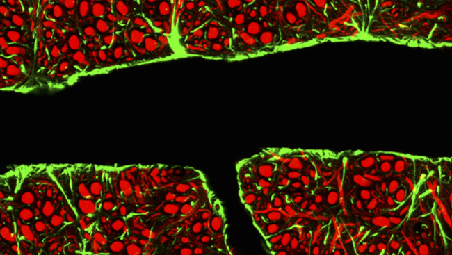
The brain links smells and sensations to create lasting memories.

Campbell, et al. The Journal of Neuroscience, 2013.
The Mushroom Body
Sex Differences in the Brain
His and Hers: Sex Differences in the Brain

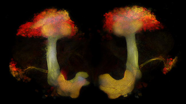
Studying sex differences in the brain may one day lead to new information about brain illnesses that affect one sex more than the other.

Shutterstock.com
Sex Differences in the Brain
scientists labeled a transcription factor called Fezf2 (in green) in cells in the motor area of the mature mouse cortex. Neurons in the layer above (in red and blue) did not express Fezf2. The gray cells show how the shape of the neurons change after Fezf2 is expressed, growing tufted dendrites.
Image of the Week: Forming a New Identity

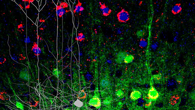
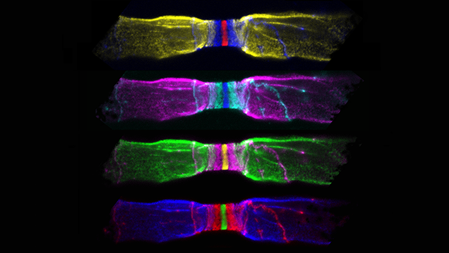
Neurons rely on proteins to determine their jobs in the nervous system.

Tantirigama, et al. Journal of Neuroscience, 2014.
scientists labeled a transcription factor called Fezf2 (in green) in cells in the motor area of the mature mouse cortex. Neurons in the layer above (in red and blue) did not express Fezf2. The gray cells show how the shape of the neurons change after Fezf2 is expressed, growing tufted dendrites.
Image of man playing the trumpet.
Does practice make perfect?

While experts debate the type and length of practice that is optimal for success, one thing is clear: training improves performance and changes the brain.

Courtesy, with permission: Carlos Delgado.
Image of man playing the trumpet.
Olfactory bulbs
Image of the Week: Seeing Your Sense of Smell

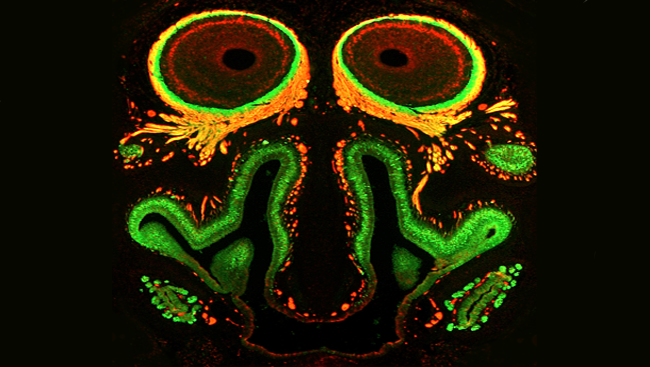
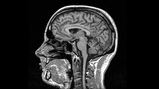
This visage captures the parts of the nose and brain responsible for interpreting smell.

Memi, et al. Journal of Neuroscience, 2013.
Olfactory bulbs
The brain’s blood vessels are lined with endothelial cells that are wedged tightly together, creating a nearly impermeable boundary between the brain and bloodstream. This image shows a section through a blood vessel (black) in the brain of a mouse as well as endothelial cells (surrounded by glial cells in green) and processed from surrounding brain cells (in red).
The Blood-Brain Barrier

Identifying new ways to bypass the brain’s elaborate security system may one day lead to better outcomes for patients with brain tumors or other neurological disorders.

Credit: C.J. Guerin, MRC Toxicology Unit / Science Source.
The brain’s blood vessels are lined with endothelial cells that are wedged tightly together, creating a nearly impermeable boundary between the brain and bloodstream. This image shows a section through a blood vessel (black) in the brain of a mouse as well as endothelial cells (surrounded by glial cells in green) and processed from surrounding brain cells (in red).
Myelin — a fatty material that wraps around axons (pictured on the left and right of nerve fibers above) — preserves the strength of these signals, allowing them to move quickly along the axon. At each node, or gap in the myelin sheath (center above), electrical signals are amplified, allowing the signal to jump to the next node.
Image of the Week: Speeding Up Brain Signals

Your brain’s electrical signals travel from node to node in their journey along nerve axons.

Desmazieres, et al. Journal of Neuroscience, 2014.
Myelin — a fatty material that wraps around axons (pictured on the left and right of nerve fibers above) — preserves the strength of these signals, allowing them to move quickly along the axon. At each node, or gap in the myelin sheath (center above), electrical signals are amplified, allowing the signal to jump to the next node.
MRI scan
Brain Scans: Technologies That Peer Inside Your Head

Advances in chemistry, physics, and computer science have revolutionized neuroscience by giving scientists greater access to the living brain.

Courtesy, with permission: Oxford Centre for Functional MRI of the Brain, Oxford University.
MRI scan

It’s been a big year for the brain and for BrainFacts.org. We published dozens of articles about the brain and nervous system, covering everything from hungry fruit bats to the eerie sensation of déjà vu. What were the most popular posts of 2014? Browse through the images and their descriptions to find out, and click on the link in each caption to read the full articles.

CONTENT PROVIDED BY

BrainFacts/SfN