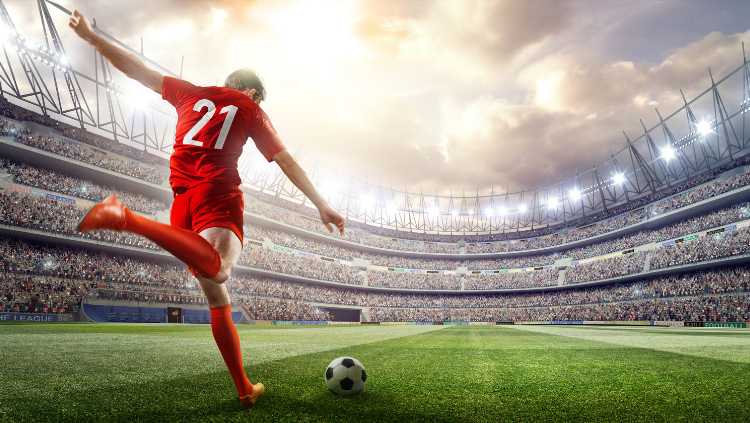
Performing Complex Movements
- Reviewed5 Oct 2022
- Author Karen Hopkin
- Source BrainFacts/SfN
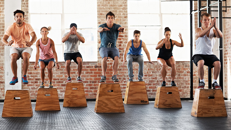
Movement is a cooperative. From swinging a baseball bat to waving at a friend across the street, movement goes beyond the involvement of muscles — multiple components of the nervous system participate in the process.
Spinal circuits, nerves extending from the spinal cord to muscle groups, play a critical role in controlling more sophisticated, voluntary behaviors, such as the alternating action of the legs during walking. In fact, the rhythmic patterns of muscle activation that produce locomotion — not only in four-footed animals, but in humans — are generated by neurons within spinal cord and brainstem circuits. When these neuronal circuits (central pattern generators) are activated, they produce the rhythmic patterns that occur in walking, flying, swimming, or breathing. Central pattern generators which evolved in primitive vertebrates are being studied to determine the degree to which spinal circuitry can be co-opted to recover basic postural and locomotor function after severe paralysis.
The most complex movements that you perform, including those requiring conscious planning, involve input from the brain. These higher brain regions initiate voluntary motion, coordinate complex sequences of movement, and tailor behavioral output to suit a given situation. Successful execution of these programs requires your brain to relay commands to the appropriate spinal circuits.
Through careful animal experiments, scientists are just beginning to understand the coordinated series of interactions that take place among different brain regions during voluntary movement. One brain area essential for voluntary movement is the motor cortex. Neurons in the motor cortex send signals that directly control the activation of alpha motor neurons in the spine. Some of these cortical neurons control the movement of functionally related muscles in an individual body part, such as your hand or arm; such neurons are important for finely tuned motor skills. Other neurons in the motor cortex can direct the coordinated movement of a limb to a particular point in space — raising your arm in a defensive position or bringing a hand to your mouth to deliver a tasty morsel of food.
Regions that Modulate Voluntary Movement
The motor cortex does not act alone in controlling complex or skilled voluntary movements. Several other brain regions participate in parallel circuits or “loops” to modulate motor control. These regions — including the basal ganglia, thalamus, cerebellum, and a large number of neuron groups located within the midbrain and brainstem — also influence the activity of motor neurons in the spinal cord. The basal ganglia themselves encompass two separate pathways. One appears to facilitate the desired motor program while the other suppresses unwanted, competing actions. Along with the thalamus, the basal ganglia share widespread connections with motor and sensory areas of the cerebral cortex, allowing these structures to monitor and adjust motor performance.
Dysfunction of the basal ganglia can lead to serious movement disorders. People with Parkinson’s disease experience degeneration of neurons in a brain region called the substantia nigra; these neurons relay signals to the basal ganglia using the neurotransmitter dopamine, a key chemical involved in motor control. Depletion of dopamine gives rise to the hallmark symptoms of Parkinson’s: tremor, rigidity, and in some cases, akinesia, an inability to move. In contrast, individuals with Huntington’s disease often display uncontrolled jerking or twitching movements, particularly in the face and extremities. These symptoms stem from a selective loss of inhibitory neurons in the basal ganglia, which eliminates the suppression of random involuntary movements.
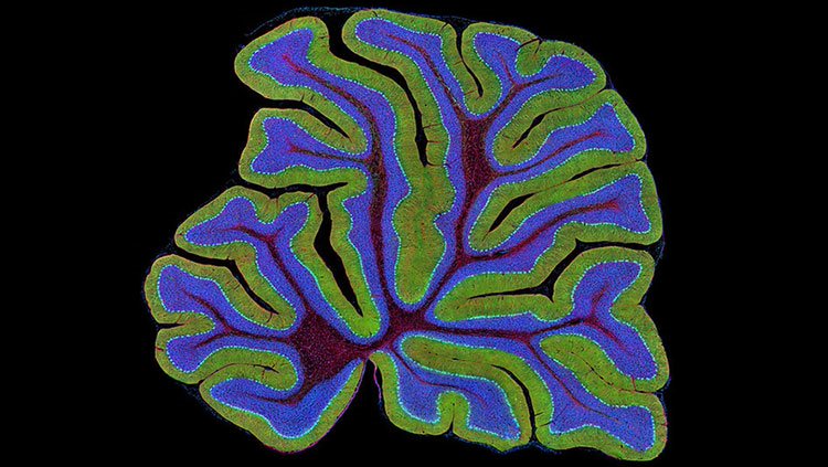
Another brain region crucial for coordinating and fine-tuning skilled movement is the cerebellum. The cerebellum receives direct input from sensory receptors in the limbs and head, as well as most areas of the cerebral cortex. Neurons in the cerebellum apparently integrate this sensory information ensuring the proper timing and integration of muscle action. This enables us to produce fluid movements more or less automatically. The cerebellum is essential to a wide range of motor learning and coordination, from controlling limb movements to eye movement to grip force.
Disturbance of cerebellar function leads to poor coordination, disorders of balance, and even difficulties in speech, one of the most intricate forms of movement control. Long-term alcohol abuse is a common cause of acquired cerebellar degeneration. Typical symptoms are poor coordination, an unsteady walk or stumbling gait, changes in speech, and difficulty with fine motor skills including eating, writing, and dressing.
The cerebellum also allows you to adapt to the unexpected, adjusting your movements so that you can smoothly lift a box that you expected to be much heavier, for example. And it plays a major role in motor learning. As you learned to walk or speak or practiced a musical instrument or a new dance routine, the cerebellum refined and sharpened the motor programs that allow you to perform these tasks with increasing accuracy and skill.
Considerable evidence also indicates that the cerebellum helps us recalibrate our movements as our own bodies change, as we grow taller, gain or lose weight or muscle mass, or cope with disease or disability. In that way, the cerebellum facilitates skillful movement through an ever-changing world as we grow up and we grow old.
Adapted from the 8th edition of Brain Facts by Karen Hopkin.
About the Author

Karen Hopkin holds a PhD in biochemistry and is co-author of the textbook Essential Cell Biology.
CONTENT PROVIDED BY
BrainFacts/SfN
References
Ataxia. 2017. Diseases and Conditions. Mayo Foundation for Medical Education and Research. https://www.mayoclinic.org/diseases-conditions/ataxia/symptoms-causes/syc-20355652
Keihn, O., Dougherty, K., Pfaff, D.W. (ed.). 2013. Neuroscience in the 21st Century, Chapter 38, Springer Science+Business Media, LLC 2013. DOI: 10.1007/978-1-4614-1997-6_42
King, S. 2010. Reflex Reactions – Our Body’s Rapid Defense Mechanism. Positive Health Online. Issue 176. Compass Internet Ltd. http://www.positivehealth.com/article/chiropractic/reflex-reactions-our-body-s-rapid-defence-mechanism
Manto, M., Bower, J.M., Conforto, A.B. et al. 2012. Consensus Paper: Roles of the Cerebellum in Motor Control—The Diversity of Ideas on Cerebellar Involvement in Movement. Cerebellum 11, 457–487 (2012). https://doi.org/10.1007/s12311-011-0331-9
Marder, E., & Bucher, D. 2001. Central pattern generators and the control of rhythmic movements. Current biology: CB, 11(23), R986–R996. https://doi.org/10.1016/s0960-9822(01)00581-4
Shanmugarajah, P.D., Hoggard, N., Currie, S. et al. Alcohol-related cerebellar degeneration: not all down to toxicity? Cerebellum ataxias 3, 17 2016. https://doi.org/10.1186/s40673-016-0055-1
Wise S.P. and Shadmehr, R. 2002. Motor Control. Encyclopedia of the Human Brain. Vol 3.