Mouse Neuromuscular Junction
- Published14 Sep 2012
- Reviewed14 Sep 2012
- Source BrainFacts/SfN
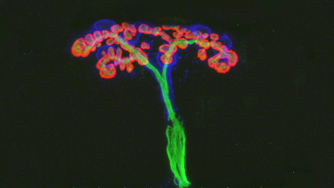
Each healthy nerve cell in the nervous system connects to other cells using limb-like structures that send and receive messages. Dendrites branch from the cell body and receive signals, while an axon extends and divides, tree-like, into smaller branches that send signals.
The image shows the end of an axon (green) connecting with a mouse’s muscle tissue. Neurons sometimes extend relatively long distances to control muscle movement. Schwann cells, glial cells found in the peripheral nervous system, are in blue. These cells make myelin, which wraps around the axon and speeds the transmission of electrical signals.
Molecules called acetylcholine receptors are clustered on the muscle fiber (red). These receptors have many subtypes. The researchers who took this image examined how specific receptor subtypes play a role in axon growth, particularly growth after injury.
CONTENT PROVIDED BY
BrainFacts/SfN