Untangling the Link Between Head Injuries and CTE
- Published16 Sep 2019
- Author Knvul Sheikh
- Source BrainFacts/SfN

The public discussion about the risks of suffering concussions, or mild traumatic brain injuries, has intensified in sports ranging from boxing and football to soccer, rugby, and ice hockey as researchers discovered chronic traumatic encephalopathy (CTE) in the brains of deceased athletes.
From ‘Punch Drunk’ Syndrome to CTE
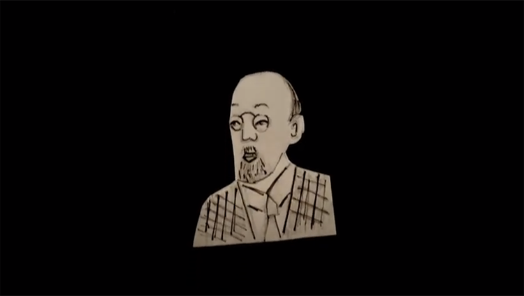
In 1928, New Jersey pathologist Harrison Martland observed boxers taking jarring hits in the ring often became ‘punch drunk’ afterward. The fighters took one or two hours to recover, but many of them developed troubling changes over time: problems with gait, tremors, and impaired memory years after they stopped fighting. The boxers most likely to suffer appeared to be the ones who spent the greatest number of years in the ring and took the most blows.
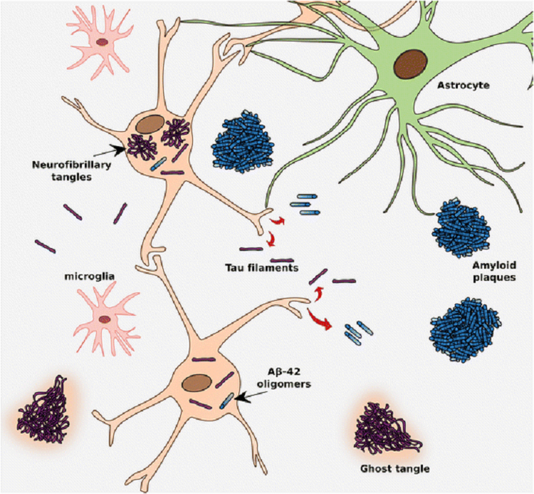
Scientists initially attributed punch drunk syndrome to the uniquely violent sport of boxing, referring to it as ‘traumatic encephalopathy of pugilists’ or ‘dementia pugilistica’ — boxer’s dementia. As researchers studied boxers, they began to identify hallmarks of the disease that would become known as chronic traumatic encephalopathy (CTE): loss of neurons in the cortex and an expansion of the brain’s ventricles — cavities in the middle of the brain where cerebrospinal fluid is produced. In addition, the boxers’ brains possessed tangled deposits of an abnormally-formed protein called tau inside neurons near blood vessels.
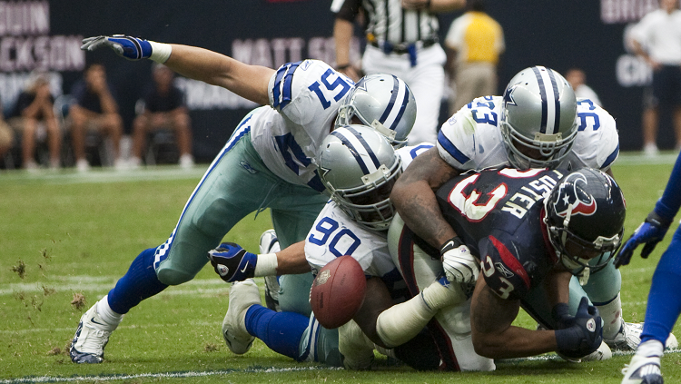
Doctors considered CTE a disease of boxers until 2005 when neuropathologist Bennet Omalu discovered abnormal tau tangles in an autopsy of American football player Mike Webster’s brain. The Pittsburgh Steelers’ legendary center had post-NFL years filled with devastating cognitive decline, depression, and erratic behavior. Webster sometimes forgot how to eat or how to find his way home. Omalu’s investigation into the source of Webster’s cognitive issues opened the floodgates for studying CTE in football players and other athletes participating in contact sports.
CTE can only be definitively diagnosed at autopsy — a tremendous hurdle for scientists and physicians hoping to treat and prevent the disease. However, during his lifetime, Webster experienced a cascade of common symptoms that people found to have died with CTE suffer from: headaches, loss of attention, and concentration. For many, their judgement can become impaired and they may suffer mood swings, depression, memory loss, difficulties with language, Parkinson’s-like tremors, and an increase in suicidal ideation.
Untangling a Mechanism
Tau is present in all of our brains. Normally, the various forms of the protein fold into soluble molecules that stabilize microtubules — structures in the neuron that act like a highway for transporting proteins from one end of the neuron to another. Tau only becomes a problem when it folds inappropriately into tangled filaments inside neurons. These neurofibrillary tangles, also called tau inclusions, arise in a number of neurodegenerative diseases including Alzheimer’s disease (AD) and CTE.
Omalu’s findings set off intense efforts to understanding how these tau inclusions form in CTE and who was at risk. In 2007, Boston University neuropathologist Ann McKee and her team made a disturbing discovery: Of 202 post-mortem brains of former high school, college, and professional football players studied, the team found CTE in 177, or nearly 90 percent of the samples. Of the former NFL players, 99 percent had CTE. That doesn’t mean 99 percent of NFL players will go on to develop CTE, but Omalu’s team established football players as a high-risk group.
“The largest risk factor for CTE that we know of right now is a history of contact sports,” says Jonathan Cherry, a neuroscientist at Boston University School of Medicine. “In fact, the more years a person plays sports or the younger they are when they start taking hits seems to speed up the progress of CTE,” he says. In one study athletes who began playing football before the age of 12 developed the cognitive and behavior symptoms of CTE 13 years earlier on average than those who started playing later in their teenage years.
With neurodegenerative diseases like AD and CTE, neural damage accumulates as tau tangles spread. The problem for scientists and physicians lies in the fact that it’s not clear how head injuries result in tau inclusions. “We really don’t know what dose of the blows to the head are needed to cause this degenerative cascade,” says Daniel Perl, a neuropathologist at the Uniformed Services University of the Health Sciences.
Developing a Diagnostic
Currently, sports teams track the impact of brain injuries by assessing players’ cognitive abilities at baseline and frequently reassessing after concussions. Such efforts may miss an important risk for CTE: subconcussions, hits which shake the brain but not so violently that there are any obvious symptoms. “Just because you don’t see an issue, because there’s no concussion or immediate unconsciousness, doesn’t mean there hasn’t been some damage to the brain,” Cherry says.
The considerable latency between suffering a blow to the head and developing symptoms associated with CTE complicates efforts to understand and address CTE, Perl says. However, scientists are beginning to get some clues about CTE thanks to technologies that weren’t available to Martland. For example, the combination of positron emission tomography (PET) and tau specific ligands allow researchers to see that there are more tau deposits in the brains of living former NFL players than other men with no history of traumatic brain injury. Whether such accumulation is predictive of developing CTE is an open question.
Michel Goedert, a neuroscientist at Cambridge University, in collaboration with Kathy Newell and Bernardino Ghetti at Indiana University, employed cryoelectromicroscopy to explore, in extremely high resolution, the structure of tau inclusions found in CTE with those found in AD. The filaments were similar but, not identical, to those found in the brains of AD patients. What’s more, Goedert’s team found CTE filaments contained a small hydrophobic molecule whereas AD does not. Previously, the researchers showed the tau filaments in Pick’s disease also differ from AD. “Our hypothesis is that the structures of tau filaments differ between diseases, but not between individuals with a given disease,” Goedert says.
Cherry, Goedert, and Perl argue the field needs concerted research efforts to develop ways to detect risk of CTE long before its devastating symptoms arise. Some research is determining if specialized PET scans will work. Other researchers are working to develop surrogate biomarkers — molecules in the blood or spinal fluid that herald the changes in the brain at the time of a blow to the head. “Developing a diagnostic test is an important first step,” Cherry says.
About the Author

Knvul Sheikh is a freelance science journalist based in New York. She writes about psychology, personalized medicine, technology and culture. Her byline has appeared in publications such as The Atlantic, Genome Magazine, Popular Science, Scholastic, Scientific American, and Vice.
CONTENT PROVIDED BY
BrainFacts/SfN
References
Falcon, B., Zivanov, J., Zhang, W., Murzin, A. G., Garringer, H. J., Vidal, R., … Scheres, S. H. W. (2019). Novel tau filament fold in chronic traumatic encephalopathy encloses hydrophobic molecules. Nature, 568(7752), 420–423. doi: 10.1038/s41586-019-1026-5
Omalu, B. I., DeKosky, S. T., Minster, R. L., Kamboh, M. I., Hamilton, R. L., & Wecht, C. H. (2005). Chronic Traumatic Encephalopathy in a National Football League Player. Neurosurgery, 57(1), 128–134. doi: 10.1227/01.NEU.0000163407.92769.ED
Mez, J., Daneshvar, D. H., Kiernan, P. T., Abdolmohammadi, B., Alvarez, V. E., Huber, B. R., … McKee, A. C. (2017). Clinicopathological Evaluation of Chronic Traumatic Encephalopathy in Players of American Football. JAMA, 318(4), 360–370. doi: 10.1001/jama.2017.8334
Stern, R. A., Adler, C. H., Chen, K., Navitsky, M., Luo, J., Dodick, D. W., … Reiman, E. M. (2019). Tau Positron-Emission Tomography in Former National Football League Players. New England Journal of Medicine, 380(18), 1716–1725. doi: 10.1056/NEJMoa1900757