Alois Alzheimer was known as the “psychiatrist with the microscope,” and he was convinced that mental illness was a disease of the brain. After one of his long-term patients died in 1906 after exhibiting strange behavior and memory loss for much of her life, and extreme dementia for her last five years, he was able to examine stained sections of her brain in a light microscope. He immediately noticed abnormal features of her brain, and recorded in meticulous drawings the numerous plaques and tangles that have come to be the diagnostic features of Alzheimer’s disease.
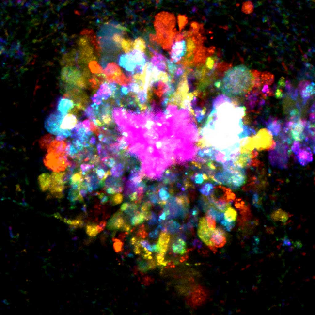
Fast forward one hundred years, and scientists are able to diagnose and attack Alzheimer’s disease from many different angles using mouse models, genetics and advanced imaging techniques. In the image above, an amyloid plaque (in purple) is surrounded by branches of damaged neurons (multiple colors) in the brain of a mouse with Alzheimer’s disease.
This article was originally published on BioInteractive.
CONTENT PROVIDED BY
BioInteractive
.jpg)