2014 Nobel Prize in Chemistry: Taking Microscopy to the Nanodimension
- Published8 Oct 2014
- Reviewed8 Oct 2014
- Author Jennifer Carr
- Source BrainFacts/SfN
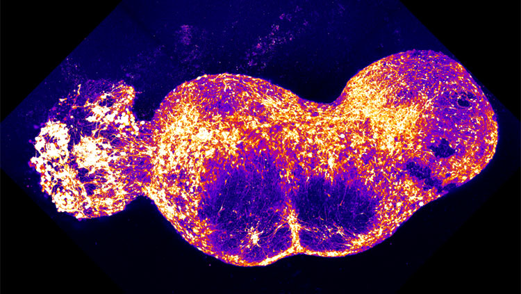
Techniques that increase the resolution of microscopes are revolutionizing the ability to peer at brain structures in ever-increasing detail, and have been awarded the 2014 Nobel Prize in Chemistry. Such technology is being used to view dendritic spines — the branchlike projections on neurons that receive inputs from other neurons — more clearly than ever before.
The top image shows the view of dendritic spines using standard confocal microscopy compared with stimulated emission depletion (STED) microscopy, one of the Nobel-winning techniques. Using STED to view living cells, scientists were able to create a 3-D image of dendritic spines from hippocampal cells in a mouse (bottom).
The ability to view molecules at the nanoscale is making it possible for scientists to see how molecules create synapses between nerve cells in the brain and track the aggregation of proteins involved in Parkinson's, Alzheimer's, and Huntington's diseases.
The Nobel Prize in Chemistry for 2014 was awarded to Eric Betzig of Janelia Research Campus, Stefan W. Hell of Max Planck Institute for Biophysical Chemistry, and William E. Moerner of Stanford University.
About the Author

Jennifer Carr is the former manager of science writing at the Society for Neuroscience. While working as a technician at a neuroscience lab at the University of Pennsylvania, Jennifer discovered she is happiest when communicating the excitement of scientific discovery to the general public. She has written for Kaiser Health News, The Scientist, and The Times-Picayune.
CONTENT PROVIDED BY
BrainFacts/SfN
References
Nagerl UV, Bonhoeffer T. Imaging Living Synapses at the Nanoscale by STED Microscopy. The Journal of Neuroscience. 30(28): 9341-9346 (2010).
Press Release. Royal Swedish Academy of Science. October 8, 2014.