Mapping and Modulating the Human Brain
- Published24 Jun 2025
- Author Sarah Heilbronner
- Source BrainFacts/SfN
Neurons are built for communication. As specialized brain cells passing signals between one another, neurons drive our every thought, feeling, and action. They cannot send messages anywhere — neurons must be physically connected to communicate.
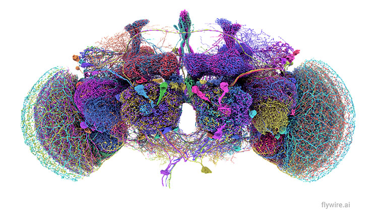
Those connections take the form of long, slender extensions on neurons which carry signals much like internal wiring in electronic devices. These extensions, called axons, are so vast that they fill over half the brain’s total volume.
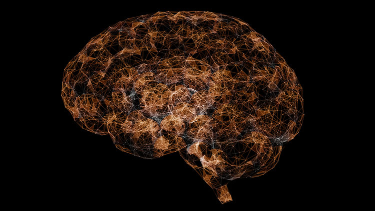
Understanding brain functions requires us to know how neurons connect to each other. Mapping the brain’s connections could quickly transform research, medicine, and mental health care. But we still do not have a full wiring diagram of the human brain.
Our most accurate method for mapping brain connections can only be used in animals.
So, why do we know so much about the brain but still don’t have a map of how everything connects? The problem is not a lack of interest, but a lack of technology. Our most accurate method for mapping brain connections can only be used in animals. It involves injecting dye into the brain, removing it, and examining it under a microscope. Obviously, we cannot do this in humans. While animal studies give us valuable insights, human brains are not the same and — more importantly — human behavior is not the same.
To close this technological gap, the United States National Institutes of Health launched an ambitious program called BRAIN CONNECTivity across Scales, or BRAIN CONNECTS, in September 2023. Part of the broader BRAIN Initiative, it seeks to tackle one of neuroscience’s most difficult challenges: mapping how brain cells are wired together. As part of the BRAIN CONNECTS consortium, my colleagues and I are working to create accurate wiring diagrams of the human brain, a goal that seemed out of reach not long ago.
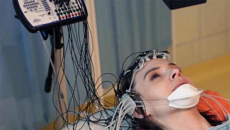
Axons form the brain’s wiring, but we cannot directly see these connections in living people. Instead, we use a workaround technique called diffusion-weighted magnetic resonance imaging (dMRI), which tracks how water molecules move through brain tissue. In areas with axons, the water tends to flow along the length of those fibers rather than across them, much like hikers sticking to a trail instead of wandering through the woods. By measuring the direction of that movement in many distinct parts of the brain, we can begin to infer the paths axons take. Even though we cannot see the wires themselves, we can map out their layout by following the footprints left behind.
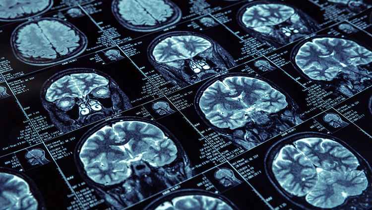
When we study the human brain after death, we have access to tools that can reveal much more detail. Thanks to the generosity of brain donors, researchers can examine brain tissue at extremely high resolution and begin tracing the paths of individual axons. One method, called polarization microscopy, takes advantage of how myelin (the fatty coating around axons) bends light in unique ways, making it easier to follow each trail through the dense landscape of the brain.
A clearer picture of how neurons connect will advance everything from artificial intelligence to cognitive neuroscience and medicine.
We are currently developing even more techniques. Plus, some advanced X-ray technologies may allow us to trace single axons. We are also improving the ways we process and label brain tissue, which may soon allow us to trace individual pathways from beginning to end. While these postmortem methods are not yet able to cover the entire brain at once, they have already been successful in smaller regions. We aim to scale them up to build a complete reference map, like a trail atlas of the human brain. By comparing this detailed map to what we see in living brains through MRI, we may one day be able to outline each person’s unique brain wiring.
A clearer picture of how neurons connect will advance everything from artificial intelligence to cognitive neuroscience and medicine. For researchers, this knowledge offers new insight into how the brain functions. However, perhaps the greatest impact will be in clinical neuroscience, which could transform how we diagnose and treat mental health conditions.
We have long known that mental health conditions are rarely caused by one fractured part of the brain. Instead, they often involve problems with how different brain areas connect and talk to each other. These connection issues are different depending on the disorder. In depression, for example, emotional areas of the brain can become too strongly linked to areas involved in self-focus and memory, while connections to areas responsible for decision-making and attention become too weak. In other words, the brain’s signals start taking the wrong paths, or missing important ones entirely.
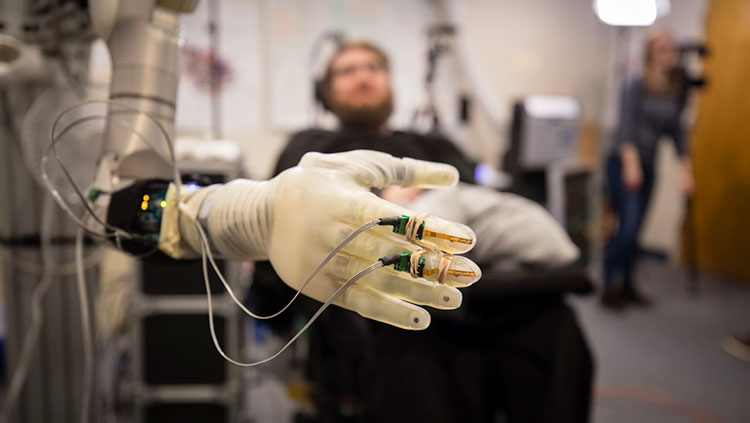
Fortunately, we already have tools to help fix these misrouted signals. Neuromodulation therapies are among some of the most promising in neuroscience. Deep brain stimulation (DBS) uses implanted electrodes to send tiny pulses into specific brain areas. Transcranial magnetic stimulation (TMS) uses magnetic fields outside the head to gently activate parts of the brain without surgery. Transcranial current stimulation (tDCS/tACS) sends small electrical currents through the scalp to guide brain activity. Focused ultrasound uses sound waves to reach deep areas of the brain with precision, also without surgery.
Right now, we use broad estimates of how the brain is wired to guide neuromodulation. But accurate, personalized maps could allow us to target them much more precisely. As our knowledge of brain connections grows, these treatments will continue to become more effective, longer lasting, and available to more people.
About the Author

Sarah Heilbronner is an Associate Professor of Neurosurgery at Baylor College of Medicine. Her lab is using cutting-edge neuroanatomical and neuroimaging techniques to try to uncover the brain’s ‘wiring diagram,’ the connections between neurons.
CONTENT PROVIDED BY
BrainFacts/SfN