This Mini Cellular Circuit Mimics Human Pain Pathway in a Petri Dish
- Published22 Aug 2025
- Author Bella Isaacs-Thomas
- Source BrainFacts/SfN
Researchers at Stanford cultivated a cellular circuit in a dish representing the neuronal activity of a pain-signaling pathway from the body to the brain.
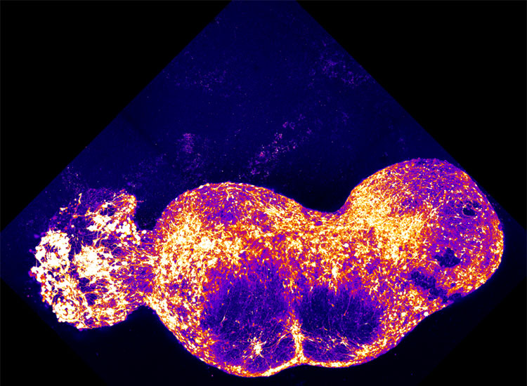
Each of the four distinct, yet interconnected, bulbs in the image above is an organoid containing around one million cells. Starting as pluripotent stem cells, or human cells capable of developing into many different cell types, these organoids arose when researchers chemically manipulated them to develop into cell clusters representing four key regions involved in pain signaling (left to right): the dorsal root ganglion (sensory neurons), the dorsal spinal cord, the thalamus, and the somatosensory cortex.
When researchers placed the four organoids beside each other in the order pain signals travel in the human pain sensory pathway, they fused to form an assembloid, or an assembly of organoids, in around 100 days. The assembloid began to demonstrate coordinated neuronal activity without intervention, according to the Stanford Medicine News Center. Researchers applied capsaicin, the chemical which causes the burning sensation we feel when we eat spicy peppers, successfully triggering activity across the assembloid.
While this assembloid does not represent all the brain and bodily regions involved in experiencing pain, these super small cells can help researchers study pain disorders. In a video, Stanford neuroscientist Sergiu Pașca said this circuit could also serve as a model researchers can use to fast track testing the efficacy of novel pain medications and more quickly offer new options to the people who need them.
About the Author
.jpg)
Bella is the Staff Writer and Editor for BrainFacts. She previously worked as a digital science reporter at PBS NewsHour, where she was named a fellow in the 2022-2023 class of the National Science-Health-Environment Reporting Fellowships (SHERF) and earned a 2021 D.C. Science Writers Association Newsbrief Award for writing. She graduated from the University of Michigan with a bachelor's degree in 2018.
CONTENT PROVIDED BY
BrainFacts/SfN
References
Kim, J. I., Imaizumi, K., Thete, M. V., Hudacova, Z., Jurjuţ, O., Amin, N. D., Scherrer, G., & Paşca, S. P. (2024). Human assembloid model of the ascending neural sensory pathway. bioRxiv, 2024.03.11.584539. https://doi.org/10.1101/2024.03.11.584539
Kim, J. I., Imaizumi, K., Jurjuț, O., Kelley, K. W., Wang, D., Thete, M. V., Hudacova, Z., Amin, N. D., Levy, R. J., Scherrer, G., & Pașca, S. P. (2025). Human assembloid model of the ascending neural sensory pathway. Nature, 642(8066), 143–153. https://doi.org/10.1038/s41586-025-08808-3