Face Blindness: How the Brain Perceives — and Sometimes Fails to Perceive — Human Faces
- Published4 Dec 2013
- Reviewed4 Dec 2013
- Author Michael W. Richardson
- Source BrainFacts/SfN
Imagine not being able to spot your loved one in a sea of faces. Or standing in front of a mirror but not recognizing the reflection staring back at you. For American artist Chuck Close and the estimated one in 50 people worldwide who live with prosopagnosia, also known as face blindness, such scenarios can be a reality. This composite image is used in tests for prosopagnosia, also known as face blindness.
During the 2012 Dialogues between Neuroscience and Society lecture, held each year to explore the intersection between neuroscience and everyday life, Close explained that he has refused to allow his face blindness and other neurological conditions to keep him from creating the gigantic portraits that have made him famous.
While some people fail to develop the ability to recognize faces, others, like Close, experience injury or disease that makes previously familiar faces suddenly unknown. Close can see fine, but cannot remember faces he has seen before.
Scientists are interested in determining how the brains of people with face blindness differ from those of healthy people so they can better understand how the brain extracts key information from the faces of others. They hope such information will shed light on this disorder, which often leads to recurrent and negative social interactions for those affected.
About Face Recognition
Face perception plays a vital role in the social interactions of humans as well as other species. In addition to serving as a primary means of recognition, studies show people have evolved to detect information about mood, focus of attention, and other cues from the faces of others.
Numerous studies of healthy people suggest the brain processes faces differently than other visual information. For example, people have a harder time recognizing or remembering faces when they are displayed upside down than they do recognizing other objects displayed upside down. Other research finds people more accurately discern parts of faces, such as a nose, when they are presented in the context of the whole face than when presented in isolation. When people are asked to discern between the features of an object, such as the windows of a house, however, they do not need to see the entire house. Studies of people with prosopagnosia, who are often able to recognize objects, also suggest the brain uses distinct mechanisms to recognize faces.
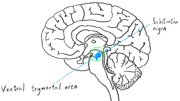
Neuroimaging studies have pointed to regions of the brain that appear to be selectively involved in the perception of faces. When adults view faces, for example, a network of regions in the occipital and temporal lobes of the brain become more active than when viewing objects.
The Search for New Clues about Face Processing
While damage to the occipital and temporal lobes is often associated with the onset of prosopagnosia, scientists are increasingly interested in the nature of the deficit in people who are face blind due to issues that arise in development.
Scientists hope that studying people with developmental prosopagnosia will lead to new insights about the mechanisms underlying face and non-face processing. For instance, early studies show people with developmental prosopagnosia tend to have family members who also struggle with face processing, suggesting there may be a genetic component to the disorder.
There is currently no treatment for prosopagnosia, and people who are face blind often learn to compensate by focusing attention on other features, such as a person’s hair style or body shape. For Close, who has no memory for a face in three-dimensions, coping with the disorder in his everyday work requires he flatten the three-dimensional face into a two-dimensional photograph that he then divides into grids. The strategy enables him to commit the face to memory so that he can paint the portrait in what has now become his signature style.
About the Author

Michael W. Richardson is a writer and editor based in Brooklyn, New York, covering topics ranging from the brain and behavior to the environment.
CONTENT PROVIDED BY
BrainFacts/SfN
References
Johnson MH. Subcortical face processing. Nature Reviews Neuroscience. 6: 766–774 (2005).
Kanwisher N. Domain specificity in face perception. Nature Neuroscience. 3(8): 759 - 763 (2000).
Palmeri TJ, Gauthier I. Visual object understanding. Nature Reviews Neuroscience. 5: 291-303 (2004).
Parr L. The evolution of face processing in primates. Philosophical Transactions of the Royal Society B: Biological Sciences. 366(1571): 1764–1777 (2011).
Yin RK. Looking at upside-down faces. Journal of Experimental Psychology. 81: 141–145 (1969).