Before You Had Eyes
- Published17 Mar 2020
- Author Charlie Wood
- Source BrainFacts/SfN
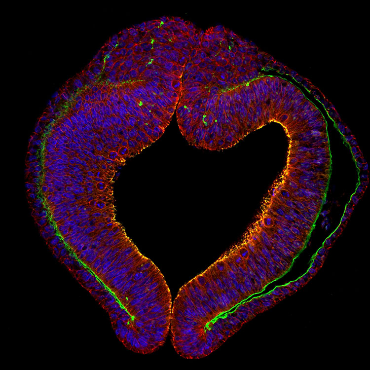
Before you had eyes, before you even really had a brain, you had a hole that your eyes and brain would grow to fill. Early in development — three to four weeks in humans, and around three days in chick embryos like the one above — two bulges start to press upward and outward from what will become the organism’s head. The bulges, or optic vesicles, will grow into an empty pathway where the brain lays wiring that becomes the optic nerve.
The tip of each bulge will soon start to pinch inward, hollowing out an early eye socket. In this image, where the right optic vesicle pokes farther into the purple region, the orange stained cells at the upper right boundary of the embryo are poised to develop into the chick’s retina — the light sensing cells that live at the back of the eye. In the rare case that a genetic condition causes this process to go awry, the organism can be born with missing or abnormally small eyes.
About the Author

Charlie Wood is a science writer with a bachelor’s degree in physics from Brown University and a master’s degree in science journalism from New York University. In previous lives he taught physics in Mozambique and English in Japan, but these days he freelances from his home in New York.
CONTENT PROVIDED BY
BrainFacts/SfN
References
Book - Text-Book of Embryology 18. (n.d.). Retrieved from https://embryology.med.unsw.edu.au/embryology/index.php/Book_-_Text-Book_of_Embryology_18.
Fuhrmann, S. (2010). Eye Morphogenesis and Patterning of the Optic Vesicle. Current Topics in Developmental Biology Invertebrate and Vertebrate Eye Development, 61–84. doi: 10.1016/b978-0-12-385044-7.00003-5