How ‘One Little Pink Dot’ Marked the Spot of the Brain’s Face-Detecting Center
- Published29 Sep 2022
- Author Juliet M. Beverly
- Source BrainFacts/SfN

She was the perfect subject to be in an fMRI scanner: she wouldn't move around, wouldn't fall asleep, wouldn't complain, but most importantly she was motivated. In 1995, neuroscientist Nancy Kanwisher was placed in an fMRI scanner at Massachusetts General Hospital in Charlestown by her research team comprised of then-undergraduate Josh McDermott and then-postdoc Marvin Chun. Before this moment, her research focused on how objects and shapes are perceived in the brain, but it would be faces that delivered her ‘Eureka’ moment.
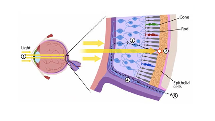
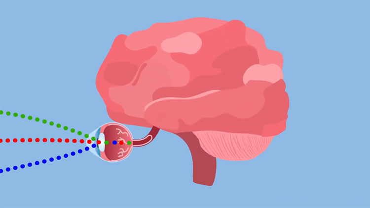
Functional magnetic resonance imaging (fMRI) relies on a person lying perfectly still while performing a cognitive task — like watching a video. The machine measures and records the changes in blood flow arising from brain activity. Kanwisher and her team deployed fMRI to understand how the brain produces the images of the objects and the environment in your field of vision. While the photoreceptors dotting the retina in the back of your eye translate the intake of light into electrical signals to relay to your brain’s primary visual cortex, it’s not a simple system. There are layers to this network of cells and tissues all over the brain, informing what you see. While the visual system helps us detect and identify objects, a debate simmered over whether a specific face-detecting area of the brain even existed in humans. Kanwisher’s study, and others, tipped the scales in a growing amount of evidence from various parts of the field.
‘There It Is’
“After I got out of the scanner, the three of us went in there [the control room] and typed away into the basic data analysis,” said Kanwisher, the Walter A. Rosenblith Professor of Cognitive Neuroscience at Massachusetts Institute of Technology (MIT). “And it was pretty fast, like a pop — this image of my brain, a whole bunch of [image] slices through my brain with this one little pink dot.” The ‘little pink dot’ highlighted an area in the brain’s inferior temporal cortex (IT). Named the right fusiform gyrus, it processes places, objects, people, and memory.
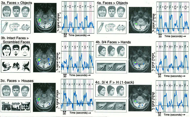
In these initial, pre-study scans, McDermott, Chun, and Kanwisher saw her response to faces, observing that the signals from particular volumes of the brain, or voxels, were higher when she viewed faces than other objects. Kanwisher's team repeated this five-minute experiment over and over, noting the same voxels in the exact same space of the brain every time a face was viewed. “You could just see big peaks during the face periods, little peaks for the objects, [then] big peaks for the face. You just see it in the data. It’s like, ‘there it is,’” said Kanwisher of the face-selective patch.
In their 1997 study, 12 of the 15 participants demonstrated increased activity within this small patch in the fusiform gyrus when they viewed gray-scale images of faces, as compared to other images shown, like houses, telephones, and animals. Their findings supported the earlier work of many researchers who found single cells specialized for perceiving hands and faces in the IT cortex. For example, in 1972 the late Charles Gross demonstrated as much with macroelectrode recordings in macaques.
Notably, the late Justine Sergent observed in a 1992 positron emission tomography (PET) study, a distinct difference in how the brain recognizes faces versus other objects — describing an indication of blood flow to the same location Kanwisher and team would later coin as the fusiform face area (FFA). The culmination of this work, and other transformative studies turned the dial from animal model to human model. “Every mental process requires many different brain regions, and face recognition is no exception,” says Kanwisher. “The thing about the FFA is not that it's the only thing we need for face recognition, but rather all it seems to do is face recognition.”
‘One Little Pink Dot’
More than 25 years after the FFA discovery, Kanwisher was able to pick at this particular thread. Working with 15 congenitally blind participants, Kanwisher and colleagues aimed to determine if visual experience was required to have this same activity in the FFA, just as she did when she first laid in the fMRI scanner in the 90s. But this time, the team prepared to work with study participants who had to move around slightly. To study the brains of blind people who never had a visual experience, the team created 3D printed objects to feel at random while in the fMRI — the tactile experience of her earlier work.
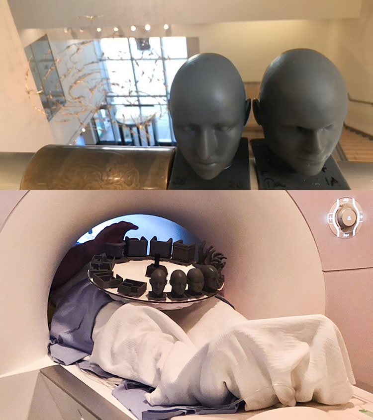
The team found the FFA responded to faces despite the patient’s having no prior visual experience of faces, showing activity in the same location as sighted patients who also handled the 3D objects. “The question of nature versus nurture — what you have already built inside your brain versus what you gain during the course of development — is a fundamental question that people have thought about in many different ways,” says N. Apurva Ratan Murty, lead author of the study and a cognitive computational research scientist working with Kanwisher and Jim DiCarlo at MIT. “In our small way, we are providing evidence that there is a part of the brain that comes online without any particular visual experience.”
The study was supported by National Institutes of Health (NIH), the National Eye Institutes (NEI), and the National Science Foundation (NSF), among others. Using experimental data from studies, Ratan Murty, who is supported by NIH’s K99/R00 Pathway to Independence Award from NEI, builds computational models of the developing human and non-human primate visual system. Knowing how much ready-made architecture should be provided in computer simulation, as opposed to how much experience, or learning, should be provided in that architecture, helps neuroscientists identify how objects are represented in the brain.
“I think it was [Richard] Feynman who very famously said, ‘What I cannot create, I do not understand,’” says Ratan Murty quoting the 1965 Nobel-winning physicist. Murty says that building a computational model of the visual system helps to eliminate human bias and bridge established theories of visual perception to more predictive models, potentially opening up new understandings into human cognition.
About the Author

Juliet M. Beverly is the senior editor for BrainFacts. She previously worked at the Embassy of Austria in the Office of Science & Technology as the assistant editor for Bridges, an online magazine covering transatlantic science and technology policy.
CONTENT PROVIDED BY
BrainFacts/SfN
References
Desimone R. Face-selective cells in the temporal cortex of monkeys. J Cogn Neurosci. 1991 Winter;3(1):1-8. doi: 10.1162/jocn.1991.3.1.1. PMID: 23964801.
Gerstein, G. L., Gross, C. G., & Weinstein, M. (1968). Inferotemporal evoked potentials during visual discrimination performance by monkeys. Journal of Comparative and Physiological Psychology, 65(3, Pt.1), 526–528. doi:10.1037/h0025813
Gross CG, Rocha-Miranda CE, Bender DB. Visual properties of neurons in inferotemporal cortex of the Macaque. J Neurophysiol. 1972;35(1):96-111. doi:10.1152/jn.1972.35.1.96
Kanwisher, N. (2017). The quest for the FFA and where it led. The Journal of Neuroscience, 37(5), 1056 1061. https://doi.org/10.1523/jneurosci.1706-16.2016
Kanwisher, N., Mcdermott, J., & Chun, M. M. (1997). The Fusiform Face Area: A Module in Human Extrastriate Cortex Specialized for Face Perception. The Journal of Neuroscience, 17(11), 4302-4311. doi:10.1523/jneurosci.17-11-04302.1997
Pinsk, M. A., Arcaro, M., Weiner, K. S., Kalkus, J. F., Inati, S. J., Gross, C. G., & Kastner, S. (2009). Neural Representations of Faces and Body Parts in Macaque and Human Cortex: A Comparative fMRI Study. Journal of Neurophysiology, 101(5), 2581-2600. doi:10.1152/jn.91198.2008
Ratan Murty, N.A., Bashivan, P., Abate, A. et al. Computational models of category-selective brain regions enable high-throughput tests of selectivity. Nature Communications 12, 5540 (2021). https://doi.org/10.1038/s41467-021-25409-6
Ratan Murty NA, Teng S, Beeler A, Mynick A, Kanwisher N (2020) Visual experience is not necessary for the development of face-selectivity in the lateral fusiform gyrus, Proc Natl Acad Sci (USA), https://doi.org/10.1073/pnas.2004607117
Sergent J, Ohta S, MacDonald B. Functional neuroanatomy of face and object processing. A positron emission tomography study. Brain. 1992 Feb;115 Pt 1:15-36. doi: 10.1093/brain/115.1.15. PMID: 1559150.