Beginning to See the Light
- Published27 Nov 2013
- Reviewed27 Nov 2013
- Author Michael W. Richardson
- Source BrainFacts/SfN
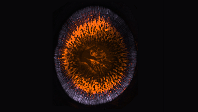
Williams, et al. Journal of Neuroscience, 2010.
The retina, a part of the nervous system that is located at the back of the eye, contains specialized cells that transmit visual information to the brain. In this image of a zebrafish retina early in development, cone photoreceptors — the cells responsible for seeing in color — are pictured in purple. Müller cells, a type of glia that provides structural stability and key support for light-sensing photoreceptors, appear in orange. Scientists observed the relationship between these two cell types during development and were surprised to find that Müller glia are not required for the cone cells to form connections. Researchers now hypothesize that photoreceptor cells are able to connect to the brain independent of glial cells, unlike other parts of the nervous system.
About the Author

Michael W. Richardson is a writer and editor based in Brooklyn, New York, covering topics ranging from the brain and behavior to the environment.
CONTENT PROVIDED BY
BrainFacts/SfN
Also In Vision


Trending
Popular articles on BrainFacts.org