Dr. Andrea Brand and Boris Egger CC BY-NC
Although a fruit fly’s brain is more than half a million times simpler than a human brain (in terms of neuron number), more than half of it is devoted to sight. After all, dodging rolled up magazines isn’t easy. These features, combined with precision modern gene techniques, make the fruit fly’s visual system one of neuroscience’s best understood models.
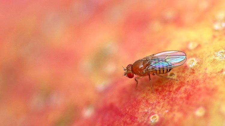
This image catches the development of that system in action. Green stem cells on the right split in such a way that they produce both a specialized neuron and a general stem cell, which is ready to split again. These neurons will link up with the green axons on the left to help carry light signals from the eye at the top.
Researchers hope to learn more about how stem cells know when to start spinning off neurons instead of continuing to split into more and more stem cells, because making new neurons would be a promising way to treat neural disorders. Neuroscientists can produce neurons in test tubes but studying the process in living animals — like flies — helps them understand how such cells might behave in people.
CONTENT PROVIDED BY
BrainFacts/SfN
References
Zhu, Y. (2013). The Drosophila visual system: From neural circuits to behavior. Cell Adhesion & Migration, 7(4), 333–344. doi: 10.4161/cam.25521
Also In Animals in Research


Trending
Popular articles on BrainFacts.org