The Pathway That Helps Know What You See
- Published6 Aug 2019
- Author Charlie Wood
- Source BrainFacts/SfN
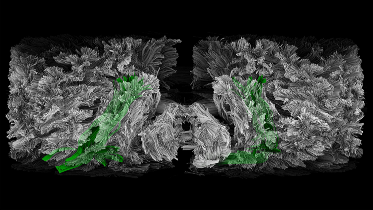
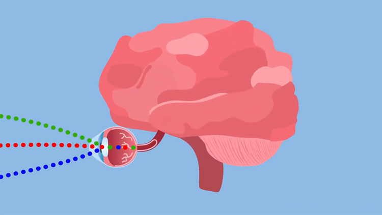
Like a cranial bigfoot, the existence of this green pathway has long been controversial. But MRI machines tracking the movement of water molecules have confirmed that the visual areas at the back of the brain do connect directly to the speech comprehension areas on either side. Researchers call this wiring the inferior longitudinal fasciculus (ILF).
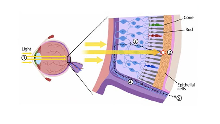
Technology has advanced enough to show that the ILF has four layers and connects with a handful of nearby pathways. Early research suggests that one of the link’s many functions is to help us understand what we see. People with damage to the right ILF have trouble recognizing familiar faces, for instance, while those with damage to the left understand less of what they read.
But IFL links appear to be plastic. One study found that studying Morse Code strengthened the ILF. Some researchers speculate that stroke patients who recover their speech do so in part by reforming this connection.
About the Author

Charlie Wood is a science writer with a bachelor’s degree in physics from Brown University and a master’s degree in science journalism from New York University. In previous lives he taught physics in Mozambique and English in Japan, but these days he freelances from his home in New York.
CONTENT PROVIDED BY
BrainFacts/SfN
References
Herbet, G., Zemmoura, I., & Duffau, H. (2018). Functional Anatomy of the Inferior Longitudinal Fasciculus: From Historical Reports to Current Hypotheses. Frontiers in Neuroanatomy, 12, 77. doi: 10.3389/fnana.2018.00077
Shinoura, N., Suzuki, Y., Tsukada, M., Katsuki, S., Yamada, R., Tabei, Y., … Yagi, K. (2007). Impairment of Inferior Longitudinal Fasciculus plays a Role in Visual Memory Disturbance. Neurocase, 13(2), 127–130. doi: 10.1080/13554790701399254
Also In Vision


Trending
Popular articles on BrainFacts.org