Light and Time: The Discovery of a New Photoreceptor System within the Eye
- Published1 Jun 2012
- Reviewed5 Jun 2012
- Source Gatsby Charitable Foundation
Until the late 1990s it seemed inconceivable to most vision biologists and ophthalmologists that there could be an unrecognised class of photoreceptor within the vertebrate eye. After all, the eye was the best understood part of the central nervous system.
Russell Foster is a professor of circadian neuroscience and head of the department of ophthalmology at Oxford University
One hundred and fifty years of research had explained how we see: Light is detected by the rods and cones and their graded potentials are assembled into an image by inner retinal neurones, which then trigger the retinal ganglion cells whose axons form the optic nerve, and after layers of neuronal processing, an image is created in the visual cortex. This representation of the eye left no room for an additional class of ocular photoreceptor. However, two parallel lines of investigation, one in fish and the other in rodents, overturned this conventional view of the eye. We now know that the rods and cones are not the only photosensory neurones of the vertebrate eye, and this discovery in humans is having a major impact on clinical ophthalmology.
Fish lead the way
The discovery of the VA-opsin gene family in fish led to the demonstration in 1998 that a sub-set of inner retinal horizontal and ganglion cells are directly light sensitive. These results provided the first unambiguous evidence for a non-rod, non-cone photoreceptor within the eye of any vertebrate and the vital “proof of principle” supporting a growing body of evidence that the mammalian eye might also contain such photoreceptors.
A curious finding in mammals?
A long-standing question had been how circadian rhythms (24h body clocks) of mammals are regulated by light. Puzzling results from a range of animal models with genetic defects of the eye showed that visual blindness and loss of most (but not all) of the rods and cones did not alter the ability of the 24h circadian clock to align (entrain) to the light and dark of dawn and dusk. Eye loss in these mice would completely block these responses to light. Although these results were consistent with the possibility that mammals might possess another light sensor within the eye, these initial studies could not preclude the alternative explanation that only a very small number of rods and/or cones mediate these effects of light. Indeed, as this author experienced, there was huge and hostile resistance initially to the very notion of non-rod, non-cone photoreceptors within the mammalian eye.
Mammals also possess novel photoreceptors
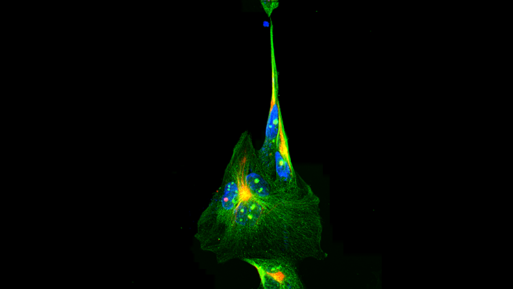
This ambiguity led to the development of a genetically-engineered mouse model entirely lacking rods and cones (rd/rd cl), and the demonstration around the turn of the century that a broad range of responses to light including the regulation of circadian rhythms, hormonal rhythms and pupil constriction all occur in the absence of the rods and cones. There had to be another photoreceptor – but what was it? Over the next five years researchers using the rd/rd cl mouse, rats, and the macaque monkey, showed that the eye contains a small number of directly light-sensitive retinal ganglion cells (pRGCs) that utilize the photopigment melanopsin (OPN4) which is maximally sensitive to blue light (λmax ~ 480nm). So, unlike fish, mammals don’t have VA opsin but another new photopigment molecule - melanopsin. However, now we know that fish have both VA opsin and melanopsin photopigments within the eye, and that most if not all the cells in the inner retina (horizontal, bipolar, amacrine and ganglion cells) can detect light!
Humans are like mice
Studies in two profoundly blind subjects with genetic diseases of the eye, and lacking functional rods and cones showed that we also have pRGC photoreceptors maximally sensitive in the blue part of the spectrum. Like mice these cells not only regulate the body clock but also sleep and pupil constriction. And remarkably, seem to provide us with a subconscious “awareness” of light. These basic findings in animal models and most recently humans are now informing clinical ophthalmology and the advice given to patients with eye diseases.
Ophthalmology has become more complicated
Until recently, body clocks and sleep timing were rarely addressed in ophthalmology and specific guidelines relating to sleep disturbance in ocular disease are lacking. It is important to stress that sleep disruption is much more than the frustration of feeling sleepy at an inappropriate time. The sustained disruption of sleep is closely linked to the added susceptibility of a range of health problems, including cognitive decline, depression and attentional failures. Thus, ocular disease not only causes visual loss but has the potential to inflict multiple additional health problems. For example, people with eye diseases of the inner retina, which result in retinal ganglion cell death (e.g. glaucoma), are at particular risk of circadian rhythm and sleep disruption. Furthermore, individuals lacking eyes entirely because of trauma will have no ability to regulate their biological rhythms. Such individuals should receive counselling regarding the problems of sleep disruption, and would be strong candidates for treatment with appropriately timed medications that help consolidate sleep timing. By contrast, eye diseases associated with rod and cone photoreceptor death need not result in the loss of pRGC photoreception. In these cases individuals should be encouraged where possible to expose their eyes to sufficient day-time light to maintain normal circadian regulation and sleep-wake timing.
Global Implications for Health
The World Health Organization suggests that at any one time worldwide 49 million people will have vision loss and over 270 million severe sight problems. There are also about 250 million people with visual impairment; it is very likely that many of these individuals could benefit from an understanding of how their visual blindness might be affecting their pRGC system, and by extension their biological rhythms of sleep and allied physiology. Unfortunately, few people worldwide are even aware of this new light sensing system and the important role it plays in regulating our physiology and behavior.
BrainFacts.org
This short essay has considered some of the recent progress in understanding the eye, but of course, every branch of neuroscience has delivered staggering and exciting innovations. Because there have been so many important developments in the neurosciences in recent years there has never been a more important time to communicate this progress to the broader non-scientific community. Not simply to take the ideas and results from the laboratory bench into clinical practice or industrial application, but also to explain to the broader community what is being done in their name and why it is so important. A failure to communicate leads to mistrust and even rejection. BrainFacts.org is playing a key role in sharing the latest developments in neuroscience with the non-specialist; exploring why today’s research is important; what we don’t yet fully understand and why science and the scientific approach belongs to us all.
CONTENT PROVIDED BY
Gatsby Charitable Foundation