The Motor Cortex
- Published20 Mar 2012
- Reviewed20 Mar 2012
- Source CIHR – Institute of Neurosciences, Mental Health and Addiction
So many different structures in the brain are involved in motor functions that some people even say that practically the entire brain contributes to body movements. Though the motor cortex is usually associated with Areas 4 and 6, the control of voluntary movements actually involves almost all areas of the neocortex.
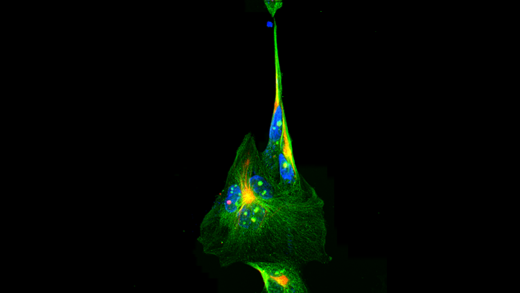
The anatomical region of the brain known as Area 4 was given the name primary motor cortex (symbol: M1) after Penfield showed that focal stimulations in this region elicited highly localized muscle contractions at various locations in the body. This mapping is represented somatotopically on the motor cortex, where the surface area devoted to controlling the movements of each body part varies in direct proportion to the precision of the movements that can be made by that part.
The motor cortex also includes Area 6, which lies rostrally to Area 4 and is divided into the premotor area (or premotor cortex) and the supplementary motor area. The premotor cortex is believed to help regulate posture by dictating an optimal position to the motor cortex for any given movement. The supplementary motor area, for its part, seems to influence the planning and initiation of movements on the basis of past experience. The mere anticipation of a movement triggers neural transmissions in the supplementary motor area.
Besides the frontal cortex, the posterior parietal cortex clearly plays a role in voluntary movements, by assessing the context in which they are being made. The parietal cortex receives somatosensory, proprioreceptive, and visual inputs, then uses them to determine such things as the positions of the body and the target in space. It thereby produces internal models of the movement to be made, prior to the involvement of the premotor and motor cortices.
Within the posterior parietal cortex, two particular areas are distinguished. Area 5 receives information from somatosensory areas 1, 2, and 3 of the cortex. Area 7 further integrates the already highly integrated signals from the visual areas of the cortex, such as MT and V5.
The parietal lobes are themselves closely interconnected with the prefrontal areas, and together these two regions represent the highest level of integration in the motor control hierarchy. It is here that the decisions are made about what action to take. The posterior parietal and prefrontal areas send their axons to Area 6 which, once it has been informed about the kind of action to take, helps to determine the characteristics of the appropriate movement for this purpose.
CONTENT PROVIDED BY
CIHR – Institute of Neurosciences, Mental Health and Addiction