‘Life of a Neuron’ Tells the Life Story of the Brain’s 86 Billion Neurons
- Published28 Sep 2021
- Author Alexis Wnuk
- Source BrainFacts/SfN
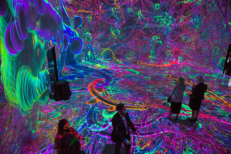
The neurons in your brain have a story to tell. Visitors to Washington, D.C.’s technology-based art space ARTECHOUSE will get a chance to watch that story unfold in Life of a Neuron.
The exhibit is the product of a years-long collaboration between ARTECHOUSE and the Society for Neuroscience, a professional organization representing neuroscientists around the globe. The immersive experience marries cutting-edge science and art to illuminate the life experience of the brain’s 86 billion neurons.
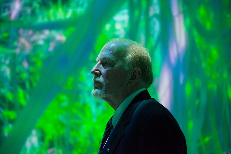
“When [people] think of the human brain … they think of this big, four-pound blob with hills and valleys,” says John Morrison, a neuroscientist at the University of California, Davis and the lead scientist behind the project. “And I want people to know that, yeah, that’s cool … but the real action is in the constant work and organization of these neurons.”
Upon descending into the high-ceilinged main room, visitors find themselves in the cerebral cortex, the wrinkled, outer layer of the brain responsible for higher cognitive functions. A looped, 20-minute animation projected on the walls and floor depicts the life cycle of the cortex’s pyramidal neurons. Lime green, egg-shaped cells radiate long fibers, and a baby can be heard babbling. That’s replaced by sounds of the schoolyard, with kids shouting and laughing and a forest of pink and purple neurons. At the end of the loop, we zoom in on an undulating pale-yellow neuron, its surface mottled like the moon. It starts to gray and stiffen, and eventually its fibers snap like aged elastic.
Constructing this animated life story involved artists using data derived from real human neurons. “I felt it had to be a human neuron because I wanted people to be able to identify with it,” Morrison says. “I wanted them to be able to say, ‘oh my god, that’s in my brain.’” But this presented the project’s biggest technical hurdle, according to Morrison. They had to combine two different microscopy techniques, each very time- and labor-intensive on their own, to build a detailed, accurate model of a human neuron.
The digital reconstruction of a three-dimensional neuron was the first step. The team used tissue samples donated from patients who had undergone brain surgery. After injecting the cells with a fluorescent dye, they slid the sample under a high-resolution microscope. But the microscope only offers a 2D glimpse. At most, you can see a tenth of micron in depth, Morrison says, and neurons occupy a space of hundreds of microns. Building the 3D model requires taking a series of thousands of images and using software to stack those images on top of each other and stitch them together.
Still, the model doesn’t reveal what’s inside the neuron. For that, the scientists turned to another technique called electron microscopy. With 100 to 1,000 times the resolution of a light microscope, electron microscopy brings a neuron’s inner world into focus – from the mitochondria powering the cell to the vesicles packaging and delivering proteins.
Morrison and his team put this internal anatomy into the first reconstruction, a technical feat that may be the first of its kind. “I’m not sure it’s been done at all,” Morrison says. Normally, scientists perform these techniques separately and extract whatever data they can from each. “The only reason we had to put it back together is because it was for visualization.”
“It took a long time,” he says. “And that’s the part that I was never sure was going to work.”
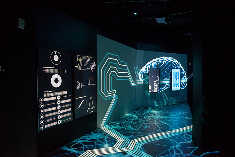
Off the main room, the wings of the exhibit house interactive installations depict the brain’s role in vision, stress, and addiction. A round screen encased in a clear dome shows the human eye while a nearby camera detects visitors as they move past it. The signal is fed to a black screen where white arrows indicate the location and direction of movement. This represents the brain’s specialized motion-detection neurons. Visual information flows from the retina, through the thalamus, and to the primary vision processing center at the back of the brain. From there, it’s divvied up and sent to neurons specialized to process shapes, edges, orientation, and motion.
"I hope that people are inspired to learn more about what makes us who we are and how we perceive the world around us."
“There’s a lot of misconceptions that vision is in the eye or that the eye is a camera,” says Ginger Leigh, the artist who created the vision installation. She hopes that her work helps people appreciate the brain’s role in vision. “I hope that people are inspired to learn more about what makes us who we are and how we perceive the world around us,” says Leigh, who is known professionally as Synthestruct. “And also realize that the way we perceive things is dictated by our brain structure and it’s not necessarily the way that things are.”
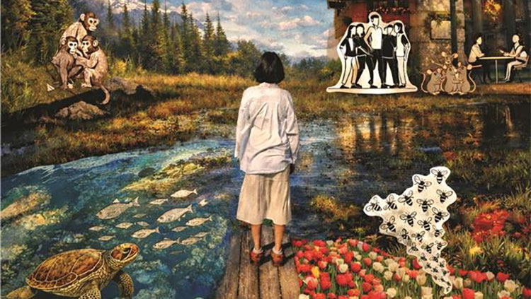
Our brains influence how we experience the world and our experience, in turn, changes our brains. The stress and addiction installations showcase the latter. Whether it’s stress or drug use, “the behavior you experience changes the neurons,” Morrison says. “Nurture is changing nature.”
Morrison also hopes the exhibit, open now through November 28, 2021, will encourage people to think of brain disorders, including those traditionally viewed as psychological, from the perspective of neurons. “That’s the only way we’re ever going to understand and treat brain disease.”
BrainFacts.org is published by the Society for Neuroscience.
About the Author

Alexis is a former staff writer/editor for BrainFacts. She graduated from the University of Pittsburgh in 2012 with degrees in neuroscience and English.
CONTENT PROVIDED BY
BrainFacts/SfN