A Journey Through the Visual System
- Published6 Nov 2014
- Reviewed6 Nov 2014
- Source BrainFacts/SfN
Explore the anatomy of the visual system as you follow light from the eye to the cortex in this video, which won People’s Choice in the 2014 Brain Awareness Video Contest. Charles Beaman, a neuroscience graduate student at the University of Texas Health Science Center at Houston, created the video.
CONTENT PROVIDED BY
BrainFacts/SfN
Transcript
We begin our journey with the eye, specifically the iris, which gives the eyes its distinctive color. Now, the iris can be green, or blue, or brown, or black depending on the level of melanin which it contains. When the lights go off, the muscles connected to the iris contract, which makes the dark circle in the center of the eye, the pupil, get bigger. When the light goes on, the pupil gets smaller to allow less light to enter. This is how the eye adapts to light.
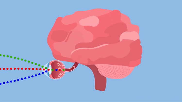
Ahh, that must be Ellie. She’ll be our tour guide on this journey through the visual system. Hi Ellie, how are you? To get a better look at the visual system, we’ll need some light. Let’s observe the anatomy of the eye in a little bit more detail. If we peel away the skin, we can see the arteries and veins, which supply important nutrients to the area. Next, we can see the surrounding muscles, which help move the eye in all directions. Now, let’s cut the eye in half to see how light enters the visual system. First, it hits the cornea, the protective layer of eye. Then, through the whole in the iris known as the pupil. Lastly, the job of the lens is to bend light to focus it correctly on the retina.
It’s time to enter the eye. Ellie has her jetpack and now we’re looking at the surface of the inside of the eye. The optic nerve on the right is where all the nerve fibers leave the eye heading toward the brain. Let’s follow Ellie has she heads toward that dark spot in the distance. This is the fovea centralis, a small pit in the retina responsible for our sharpest, clearest vision. Foveal vision allows us to do things like read books, or drive cars, or play video games.
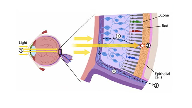
Now, let’s look at a cross-section of the retina to see how neurons respond to light. Light is absorbed by rods and cones, which are specialized photoreceptors. This starts a chain reaction, which excites the bipolar cells and then subsequently the ganglion cells, to send electrical signals off toward the brain. The amacrine and horizontal cells work to modulate the circuit.
Ok, we’ve successfully made it out of the eye, and now it’s time to head back toward the brain. Visual information flows along the optic nerve like a river of electricity. At the optic chiasm, the signals split such that images from the left visual field head to the right brain, and images from the right visual field head to the left brain.
After the optic chiasm, the visual signals make a quick stop at the lateral geniculate nucleus, or LGN. The LGN is organized into 6 layers, which all receive extensive feedback control from higher visual areas.
From the LGN, the visual signal travel along optic radiations back to the visual cortex. The cortex is where we use the signal that originally came from the eye to construct our visual reality. The billions of neurons in the human brain work to encode and process the information. Information is sent forwards and backwards. See, the beauty of the visual system is that everything we see is affected by our memories, and our feelings, and what we’ve seen before.
Well, that concludes our journey through the visual system, see you next time.