Rewiring Hope: New Insights Into Alzheimer’s and Brain Aging
- Published24 Jun 2026
- Author Jennifer Michalowski
- Source BrainFacts/SfN
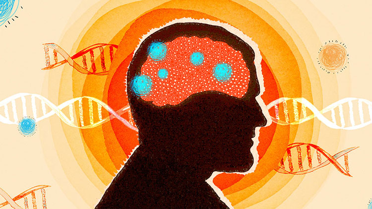
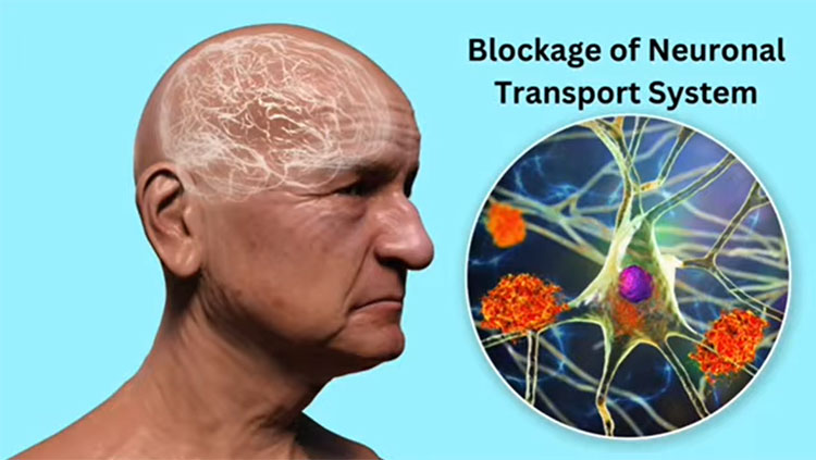
By the time someone with Alzheimer’s disease notices their memory is failing, neurons in the brain have already begun to die. Toxic proteins in the brain’s memory centers will reach further into the brain’s cerebral cortex, destroying vital connections and interfering with cognitive functions like language and reasoning. Inevitably, the disease progresses, and its spread through the brain is as predictable as it is devastating.
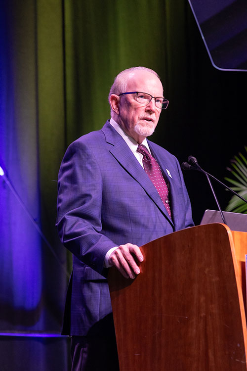
“Alzheimer's disease robs people of precisely the capabilities that are most advanced in humans,” says John Morrison, a neuroscientist at the California National Primate Research Center and past president of the Society for Neuroscience. “You can no longer think properly or think deeply about virtually anything. And of course, the memory goes as well.”
Morrison began studying Alzheimer’s disease nearly 40 years ago, intrigued by why the neural circuits we need to think, learn, and remember are the ones most vulnerable to the disease. Since then, he has clarified how age-related changes making people more forgetful later in life are distinct from the cellular destruction caused by neurodegenerative disease. Aging may set the stage for Alzheimer’s disease, but decades of studying the ways neural circuits decline have made Morrison optimistic neuroscientists will find ways to protect them.
Reversible decline
Early in Morrison’s career, little was known about why the changes observed in brains affected by Alzheimer’s disease prevented people from thinking clearly. A clearer picture emerged when his group, then at Icahn School of Medicine at Mount Sinai, and Antonio Damasio and colleagues at the University of Iowa discovered neurons connecting different parts of the cerebral cortex, where high-level cognitive processing takes place, were dying due to the disease. Without them, critical brain areas cannot work together the way they should.
Most people with Alzheimer’s disease don’t develop symptoms until their 70s or 80s. Morrison wanted to know how neurons might become more susceptible to disease as they age. Through studies in monkeys, who share similar cortical circuitry to humans, his team learned about age-related structural and molecular changes in the connections between neurons — their synapses — and how they lead to memory problems and loss of cognitive flexibility.
“At that time, people thought normal aging was mild neuron death. We spent years showing that it's actually quite different,” he says. “The circuits suffer in terms of their synaptic connections, but they don't die.”
Synaptic decline does occur early in Alzheimer’s disease, before destructive proteins begin to accumulate in and around neurons. Morrison is encouraged by this, because unlike neuron loss, synaptic decline is reversible. He suspects some additional change or exposure must provoke the transition to neurodegeneration in people who develop the disease — and it may not be the same for everyone. Inflammation, metabolic changes, or infection may play a role. One way drug developers hope to stop the disease is to block this transition, and maybe even restore synaptic health to damaged neurons.
A path toward treatment
In 2015, Morrison joined the California National Primate Research Center, where he served as director until 2024. Scientists had greatly advanced their understanding of how Alzheimer’s disease destroys neurons, but time and again, experimental drugs failed to stop the disease in patients. Researchers agreed: They needed a way to investigate potential therapies in cortical circuits resembling those in the human brain.
Morrison and colleagues made it possible by developing two primate models recreating key features of Alzheimer’s disease. One is well suited for studying the early stages of Alzheimer’s development. The other recreates Alzheimer’s progression in the brain, beginning with tangles of the protein tau in memory-associated areas like the entorhinal cortex and hippocampus, then spreading to other cortical circuits. If researchers can find ways to stop the disease’s spread, Morrison says, they might be able to limit patients’ symptoms to memory problems, preserving other cognitive functions.
Jeff Kordower, a longtime collaborator at Arizona State University who worked with Morrison to develop the tau-based model, says primate models are vital for developing Alzheimer’s therapies. “If you want to cure neurodegenerative diseases, you're not going to do it with a mouse,” Kordower says. “There are therapies that never would have taken place if the preclinical work wasn't done in monkeys.” He and Morrison are ready to work with drug developers to evaluate the effects of potential therapies in their monkey model.
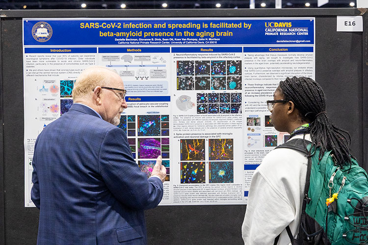
Morrison’s current outlook on Alzheimer’s research has been shaped by his role at the primate center — not just by the availability of new primate models, but also by an unexpected discovery his team made during the COVID-19 pandemic.
Studying the disease in monkeys, his group learned the virus can enter the brain through the odor-detecting circuitry that begins in the nose and quickly reaches areas where Alzheimer’s disease begins. Researchers are beginning to recognize infection and subsequent inflammation may be a significant driver of neurodegeneration. Morrison plans to investigate whether infection with the virus responsible for COVID-19 accelerates disease progression in their Alzheimer’s model and hopes more scientists will explore the intersection of neuroscience and infectious disease.
Above all, he’s convinced innovative new treatments for Alzheimer’s disease and other brain disorders are within reach, with today’s progress backed by decades of neuroscience discovery. “It all starts with the basic science,” he says. Acknowledging the uncertainty many scientists are currently facing about their labs and funding, Morrison says he remains optimistic. “It's just too important,” he says. “It's too important to let the support for it decline at all.”
About the Author

Jennifer Michalowski writes about scientists and their discoveries, exploring topics ranging from ancient evolution to the newest insights about the brain.
CONTENT PROVIDED BY
BrainFacts/SfN
References
Beckman, D., Chakrabarty, P., Ott, S., Dao, A., Zhou, E., Janssen, W. G., Donis-Cox, K., Muller, S., Kordower, J. H., & Morrison, J. H. (2021). A novel tau-based rhesus monkey model of Alzheimer's pathogenesis. Alzheimer's & Dementia, 17(6), 933–945. https://doi.org/10.1002/alz.12318
Beckman, D., Ott, S., Donis-Cox, K., Janssen, W. G., Bliss-Moreau, E., Rudebeck, P. H., Baxter, M. G., & Morrison, J. H. (2019). Oligomeric Aβ in the monkey brain impacts synaptic integrity and induces accelerated cortical aging. Proceedings of the National Academy of Sciences of the United States of America, 116(52), 26239–26246. https://doi.org/10.1073/pnas.1902301116
Duan, H., Wearne, S. L., Rocher, A. B., Macedo, A., Morrison, J. H., & Hof, P. R. (2003). Age-related dendritic and spine changes in corticocortically projecting neurons in macaque monkeys. Cerebral Cortex, 13(9), 950–961. https://doi.org/10.1093/cercor/13.9.950
Hara, Y., Rapp, P. R., & Morrison, J. H. (2012). Neuronal and morphological bases of cognitive decline in aged rhesus monkeys. Age, 34(5), 1051–1073.https://doi.org/10.1007/s11357-011-9278-5
Hof, P. R., Cox, K., & Morrison, J. H. (1990). Quantitative analysis of a vulnerable subset of pyramidal neurons in Alzheimer's disease: I. Superior frontal and inferior temporal cortex. The Journal of Comparative Neurology, 301(1), 44–54. https://doi.org/10.1002/cne.903010105
Hyman B. T., Van Hoesen G. W., Damasio A. R. Memory-related neural systems in Alzheimer's disease: an anatomic study. Neurology, 40(11):1721-1730. doi:10.1212/wnl.40.11.1721
Young, M. E., Ohm, D. T., Dumitriu, D., Rapp, P. R., & Morrison, J. H. (2014). Differential effects of aging on dendritic spines in visual cortex and prefrontal cortex of the rhesus monkey. Neuroscience, 274, 33–43. https://doi.org/10.1016/j.neuroscience.2014.05.008