Seeing the Details: What Lizard Eyes Teach Us About Vision
- Published10 Mar 2026
- Author Lesley Earl
- Source BrainFacts/SfN

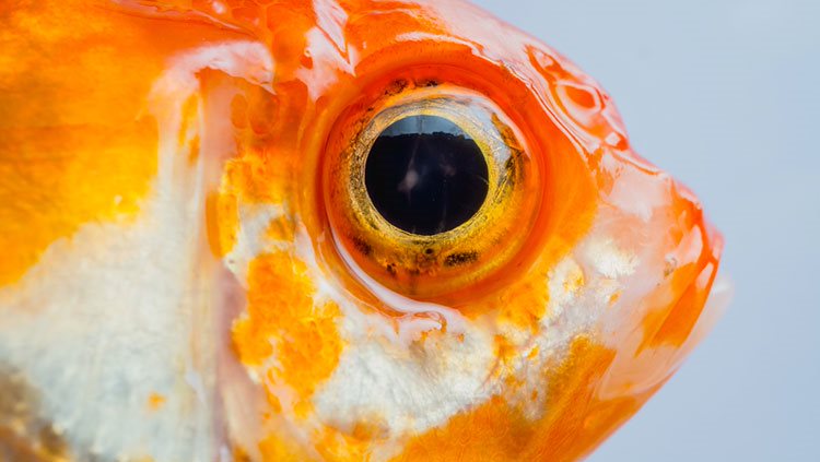
Perched silently on a thin branch, a tiny anole lizard scans its environment for prey. Catching sight of an insect with its right eye, the anole creeps into striking distance. The lizard turns its head to focus both eyes on the target, then snatches its meal with a perfectly calculated tongue flick.
For both humans and anoles, the ability to see details — such as a tasty snack — depends on a pit-like structure in the retina called a fovea, which is tightly packed with light-detecting photoreceptor neurons. While humans, and many lizards, possess just one fovea per retina, the anole has two different foveae in each eye, with distinct features and uses. Although these structures differ somewhat from the human fovea, researchers believe studying the anole visual system can teach us more about our own.
In humans, problems with foveal vision drive some of the symptoms associated with conditions like macular degeneration and ocular albinism. Issues with this part of the retina make “doing things like driving a car, reading a book, [or] being able to recognize individuals by sight at a distance very challenging,” said Andrew Wegerski, a researcher studying lizard eyes at the National Institutes of Health (NIH).
Wegerski is working on a project to develop lizards as a vision research model, a process that includes carefully imaging the cellular structure of the anole retina. Scientists hope this information could one day be used to develop and test new medications and treatments that could preserve people’s vision.
Understanding the Fovea
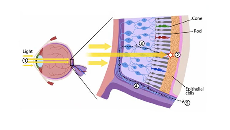
The term fovea — plural foveae — simply means “pit,” Wegerski explained. The anole’s central fovea, which it uses to assess the environment, fits that description perfectly, forming a sharp funnel shape. At the base of this funnel is a light-sensing layer of tightly packed photoreceptors, with the photoreceptors’ cell bodies and other retinal cells pushed to the side. This structure allows the anole to capture super clear images of objects positioned off to its side.
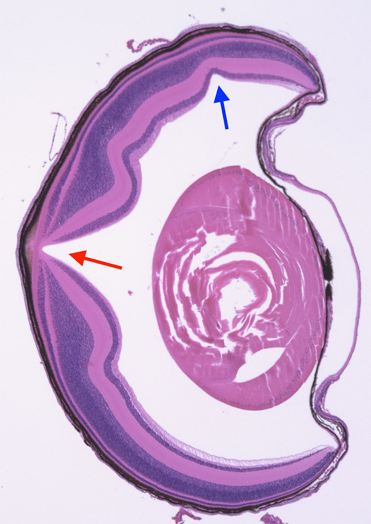
Meanwhile, the temporal fovea, which the lizards use to track and capture prey directly in front of them, is quite shallow, with only a slightly increased density of photoreceptors compared to the central fovea. The human fovea falls somewhere in the middle by comparison — it’s a modest pit with highly concentrated photoreceptors at the bottom.
Leo Fleishman, a retired researcher who studied lizard behavior and ecology at Union College, believes the anole eye evolved specifically to detect motion, allowing it to quickly sense moving objects with its deep central fovea. Other lizards, most of which don’t have a temporal fovea, rely on their central fovea for both detecting motion and gauging the distance between themselves and the moving object. But anoles, by contrast, seem to depend on the temporal fovea in the final moments of their hunt to accurately judge how far away their meal is.
The anole can use its temporal foveae to see the same object with both eyes at the same time. Humans, too, use our foveae to assess the distance of objects we see. Scientists have guessed that anoles may similarly use binocular vision — defined as such because it relies on both eyes at once — to gauge striking distance while hunting.
To investigate this phenomenon, researchers in Fleishman’s lab gently covered one anole eye as the lizards hunted. The anoles were able to close the distance to their prey, an action indicating their successful use of the central fovea. But when they attempted to capture their meal, an action that requires the temporal fovea, they missed. This outcome suggests the lizards “need both eyes” to both successfully calculate distance and catch prey, Fleishman explained.
NIH veterinarian-scientist Ashley Rasys, who works alongside Wegerski, noted a similar phenomenon in a group of albino anoles in her laboratory. Like humans with albinism, foveae in albino anoles don’t develop properly. These anoles “didn't engage in the normal hunting behavior,” and instead “just sat on their perches and waited,” Rasys said, adding “nothing seemed to catch their eye.” In fact, the lizards were so bad at hunting that researchers had to carefully hand-feed them insects.
How Vision Informs Behavior
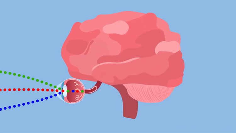
So how do healthy anoles use both eyes to judge distance? When humans look at an object, our brains automatically combine the images from each eye to give us a sense of depth. This process depends on complex neural circuitry connecting each eye to both the right and left sides of the brain; with these crisscrossing connections, the brain can mesh overlapping images from two eyes and give us a world that appears three dimensional.
Josh Morgan, a neuroscientist and microscopist at Washington University in St. Louis, studies anoles to figure out how different pieces of visual information connect in their brains. He said one can quickly get a sense of the complexity of anole visual behavior by watching their eyes when crickets are introduced into their enclosure. After detecting motion in the periphery of their visual field, they turn one eye toward a potential target so that its image falls on the central fovea. If they identify the object as prey, they then direct both eyes towards their target before pouncing. The anole’s distance detection might come from how far each eye must turn inward to maintain focus, said Fleishman. The more cross-eyed the lizard gets, the closer the object.

Understanding how neuronal connections between the eye and the brain support prey capture is one of several questions Morgan is exploring with these lizards. The anole provides a unique system “for studying the relationship between circuitry and behavior,” he said. Because different behaviors use different regions of its visual system — like prey identification with the central fovea, or navigating with the temporal fovea — the anole offers an inexpensive, attractive model to study not just eye development, but also how the brain processes vision to drive behavior.
Morgan is focusing on a part of the brain called the lateral geniculate nucleus, or LGN, a neuronal region that helps process visual information in both humans and lizards. The LGN helps us recognize visual events, like seeing a bird flying overhead, or, in a lizard’s world, judging the visual displays of another anole. Morgan hopes mapping these circuits in the lizard brain can help us better understand how our own brains function, too.
It may seem hard to believe we have much in common with the tiny anole. But Wegerski said the common ground between our vision and theirs offers us the chance to relate to an animal that otherwise appears “somewhat strange and alien.” Down the line, he added, this lizard and its unique eyes could be key to identifying new therapeutics for our own visual system: “This animal could have a very important influence.”
About the Author

Lesley Earl is a science and medical writer based in Maryland. She earned her bachelor's degree in biology from Haverford College and a Ph.D. in cellular and molecular pathology from the University of California, Los Angeles.
CONTENT PROVIDED BY
BrainFacts/SfN
References
Fleishman, L. J. (1992). The Influence of the Sensory System and the Environment on Motion Patterns in the Visual Displays of Anoline Lizards and Other Vertebrates. The American Naturalist. Vol. 139, pp. S36-S61.
Fleishman, L. J. (2024). Lizard visual ecology. Frontiers in Amphibian and Reptile Science. 2:1426675. doi: 10.3389/famrs.2024.1426675
Rasys, A. M., Pau, S. H., Irwin, K. E., Luo, S., Kim, H. Q., Wahle, M. A., Menke, D. B., & Lauderdale, J. D. (2025). Histological analysis of retinal development and remodeling in the brown anole lizard (Anolis sagrei). Journal of Anatomy, 246(6), 1019–1033. https://doi.org/10.1111/joa.14193
Wahle, M. A., Kim, H. Q., Menke, D. B., Lauderdale, J. D., & Rasys, A. M. (2023). Maturation and refinement of the maculae and foveae in the Anolis sagrei lizard. Experimental Eye Research, 234, 109611. https://doi.org/10.1016/j.exer.2023.109611
Yu, G., Katz, L. N., Quaia, C., Messinger, A., & Krauzlis, R. J. (2024). Short-latency preference for faces in primate superior colliculus depends on visual cortex. Neuron, 112(16), 2814–2822.e4. https://doi.org/10.1016/j.neuron.2024.06.005