Why Neuroscientists Are Excited for a Wearable PET Scanner
- Published9 Mar 2018
- Reviewed9 Mar 2018
- Author Rachel Kaufman
- Source BrainFacts/SfN
If you want to look at a living human brain, there are only a few ways to do it. Most of them require the owner of the brain to lay flat on their back and stay very, very still.
This limits what we can learn about our brains.
Human beings, says Julie Brefczynski-Lewis, an assistant professor of physiology and pharmacology at West Virginia University, “are very mobile. We're beings because we ... go. We talk. We gesture. We change our postures. We change our expressions.”
Not being able to do that in a scanner makes any interaction studied “a little bit artificial.”
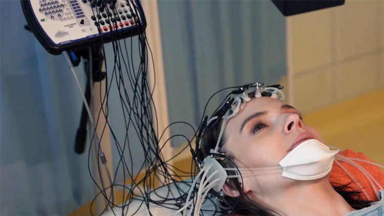
For years, neuroscientists have employed functional magnetic resonance imaging (fMRI), the gold standard of brain imaging, to track changes in the brain. The technique monitors blood flow to different areas of the brain. However, getting an fMRI scan requires a person to lie perfectly still on a bed inside an enormous cylinder. They cannot move, twitch, or sneeze. Even with these limitations, fMRI studies have advanced neuroscience and psychology for decades and in a wide variety of ways.
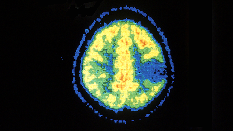
Another way to look at brain activity is via the PET scan. PET stands for positron emission tomography. This imaging technique detects the emission of subatomic particles in active areas of the brain. Researchers also use PET to study how neurotransmitters like dopamine move through the brain. Like fMRI, PET scans force people to lie perfectly still on their backs. PET scans also involve injecting people with a small amount of a radioactive tracer capable of emitting positrons, which are detected by the scanner.
Brefczynski-Lewis and her collaborator, Stan Majewski, wanted to study the brain activity of people while they were moving and employ the smallest amount of radiation possible to do so. Inspired by technology used to study laboratory rats, Brefcynski-Lewis and Majewski sketched out a solution on a piece of paper.
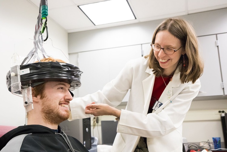
Their solution is the AM-PET — a wearable helmet that will track changes in the brain while its wearer is sitting, standing, or even moving around. The "AM" in AM-PET stands for "ambulatory micro-dose," ambulatory because the person can move around and micro-dose because the amount of radioactive substance injected is much smaller than that required for a traditional PET scan.
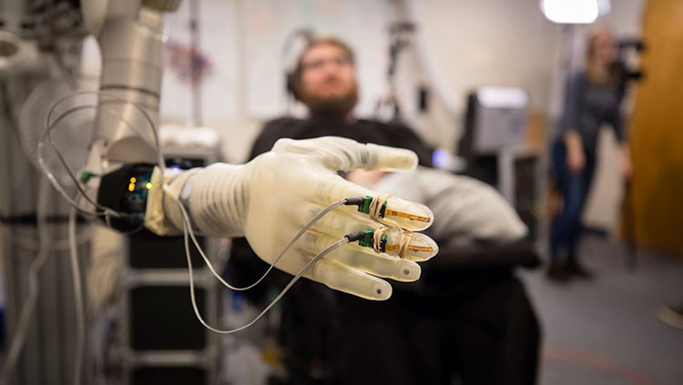
The AM-PET, which has been supported by the National Institutes of Health BRAIN Initiative, will eventually come in three versions. The first, which Brefczynski-Lewis says is one of two in testing right now, connects the scanner to a chair. The volunteer sits in the chair and wears the scanner like a salon hairdryer. The second prototype uses a harness to support the scanner from above, which means the wearer can walk in place. A third, more futuristic prototype, mounts the scanner on a robot, allowing a research volunteer to go anywhere while wearing the scanner. In the robot version, computers track the person’s head motion without ever touching it, and robotic arms realign the helmet in real time. “It’s kind of like a hovercraft for your brain,” Brefczynski-Lewis says.
Exceeding Limitations
AM-PET will allow neuroscientists to study a range of topics. “As soon as I saw this project,” says Joy Hirsch, a professor of psychiatry, comparative medicine, and neuroscience at Yale University, “I was like, ‘Oh my gosh, I’ve been so limited in terms of what I can do in terms of social neuroscience.’”
Hirsch is already envisioning ways to use the AM-PET. Her lab has focused on studying how people’s brains react as they interact with other humans. “The most important functions of the human brain are functions that relate to how we interact with each other,” she says. “We know very little about these mechanisms because they are not study-able with conventional imaging technologies.”
To date, Hirsch has employed a technology called functional near-infrared spectroscopy (fNIR) to measure signals from two people’s brains at the same time. fNIR measures the changes in cerebral blood flow that comes with brain activity. And, because it monitors blood flow with optical sensors glued to a person’s head, the technique works while people move. However, fNIR can’t measure the activity of specific neurotransmitters, such as dopamine.
Hirsch’s lab recently published a paper showing how two people’s brains sync when they make eye contact. “One can imagine that there are very important neurochemical changes associated with that,” she says, but they haven’t been studied yet.
Hirsch looks forward to the day when she can get her hands on two AM-PET scanners to explore those changes. “This eye contact business is stuff that poets have understood since the beginning of time,” she adds. “It takes neuroscientists a long time to catch up.”
In addition to helping to unlock secrets of the brain, the AM-PET helmet could have clinical implications. Swarna Rajagopalan is a neurointensivist at West Virginia University. She works at the university’s hospital in the intensive care unit, taking care of patients with severe traumatic brain injuries (TBI).
The project "has this range from the clinical to the basic research, and to what makes us human."
“Day in and day out,” she says, “I see these patients do well, or do terrible. Their scans look the same ... What is it about this person that makes them do ... so much better? The truth is, we don’t know.” It’s hard to study severe TBI patients, she says. Often their injuries are so severe that it would be dangerous for them to lie down, or they require so many medications to treat their injuries that it would be impractical to transport them to the hospital’s PET scanner. With the AM-PET, the scanner could be brought to the patient, and they would simply have to sit in a different chair.
Rajagopalan and Brefczynski-Lewis are working toward a study in which they will scan the brains of TBI patients six times, the first time within 24 hours of the patient’s being admitted to the hospital after the injury. “We can do point-of-care testing,” she says. “Not just a picture at the beginning and a picture at the end, but we can track the progress of what’s actually happening in the brain.”
The “chair” version of AM-PET is working now, though the team continues to improve it as they simultaneously seek to use it in new studies. They’re also testing the harness version on healthy people in pilot studies. This project, says Brefczynski-Lewis, “has this range from the clinical to the basic research, and to what makes us human.”
About the Author

Rachel Kaufman is a journalist covering science, technology, sustainability, and other topics. Her writing has appeared in National Geographic News, Scientific American, Inc., and more.
CONTENT PROVIDED BY
BrainFacts/SfN